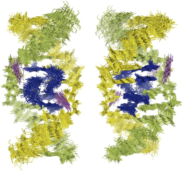
Figure 4.

Superimposed representations of the 30 lowest-energy NMR structures of the NCD-GG1 complex. Two NCD molecules are colored in blue. DNA strands are colored in yellow and green. The flipped-out cytosine bases are colored in magenta. The structures are seen from the major groove (left) and minor groove (right).
