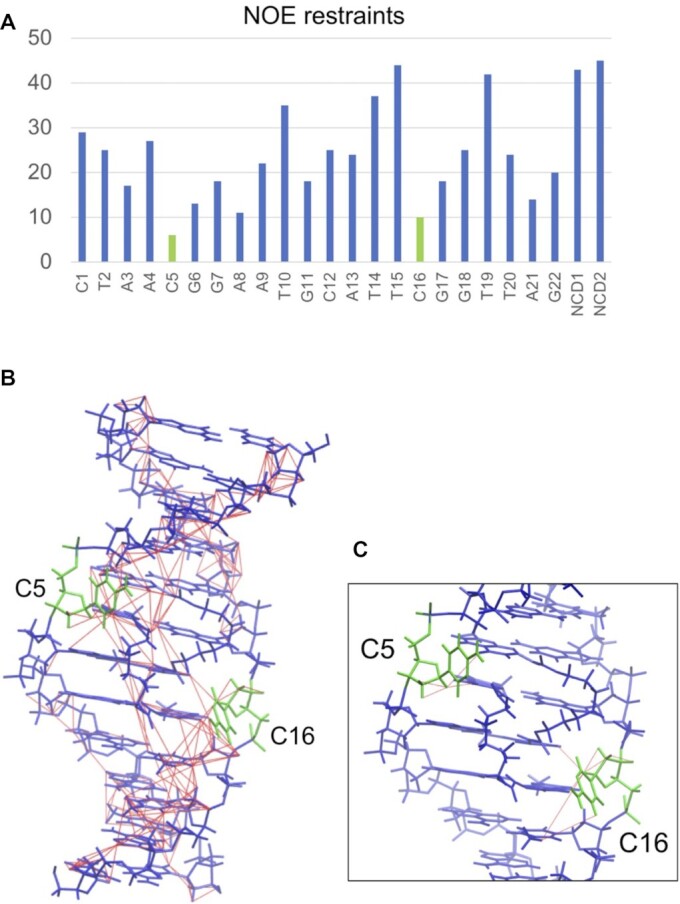
Figure 5.

(A) The number of NOE restraints for each residue. (B) NOE restraints (thin red lines, drawn by VMD-XPLOR (Schwieters, C.D. & Clore, G.M. The VMD–XPLOR visualization package for NMR structure refinement. J. Magn. Reson. 149, 239–244 (2001).)) used for the structure determination are shown on the lowest energy structure of the NCD-GG1. C5 and C16 are shown in green, and the other nucleotides and NCD molecules are shown in blue. (C) An enlarged view near C5 and C16 is shown; only NOEs containing C5 or C16 are shown.
