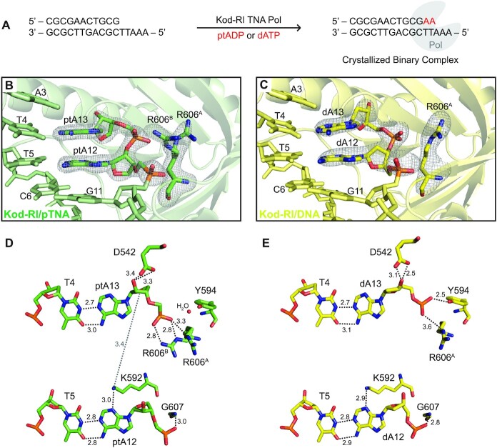
Figure 2.
Active site view of Kod-RI TNA polymerase showing pTNA incorporation. (A) Schematic representation of the crystallized binary complex following the addition and translocation of two adenosine nucleotides. (B) View of the active site of Kod-RI incorporating pTNA with a 2Fo–Fc composite polder map contoured at 7.5σ for the incorporated ptA12 and ptA13, and another polder map contoured at 5.0σ for both conformations of arginine 606, labeled A and B. The exonuclease domain is hidden to avoid obstruction of the view. (C) Kod-RI incorporating DNA with polder maps contoured at 7σ for DNA (dA12 and dA13) and 4σ for the single conformation of arginine 606. (D, E) The intermolecular interactions observed in the active site with distances reported in angstroms. Color scheme: structure obtained for the pTNA extended primer (green) and DNA extended primer (yellow).

