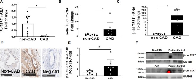
Figure 1.
Increased levels of β-del TERT expression in CAD subjects. Relative levels of FL (A), α-del (B), and β-del (C) TERT mRNA expression were measured in LV tissue from non-CAD vs. CAD subjects (N = 6–9). (D) β-del TERT expression was measured by IHC in coronary arteries isolated from non-CAD vs. CAD subjects (representative of 3 replicates). (E + F) Western blot analyses of β-del TERT levels in LV tissue of patients with and without CAD. *P < 0.05 vs. non-CAD t-test. Values are means ± SEM, N = 5–8.

