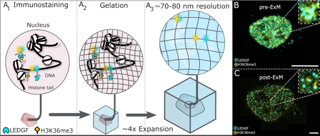
Figure 1.
ExEpi: schematic illustration of the concept and typical images obtained pre- and post-expansion. (A) Immunolabeling of the chromatin reader LEDGF and the epigenetic modification (H3K36me3) in permeabilized and fixed cells using fluorescently labelled antibodies (A1) is followed by a gelation (A2) and expansion step (A3) to improve resolution showing an actual overlap and thus interaction between LEDGF (blue) and H3K36me3 (yellow) molecules in the top, whereas the two bottom molecules no longer show co-localization in comparison to the sample before expansion. (B) Composite immunofluorescence image of the LEDGF protein in cyan and the H3K36me3 modification in yellow before gelation and expansion in the nucleus of a HeLaP4 cell. The right upper corner depicts a zoom of the boxed area. (C) Same cell as in (B) but after the gelation and expansion process with improved resolution. The presented images (B-C) are single optical sections. Scale bars: 10 μm (B, C). Details on used antibodies and dilutions can be found in SI Table 11.

