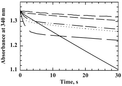
FIG. 5.
AhpC activity assay with Trx1 or Trx2 as the reductant. The decrease in NADPH absorbance was monitored on a stopped-flow spectrophotometer when AhpC (20 μM) and TrxR (0.5 μM) were assayed with Trx1 (5.0 μM, solid line) or with Trx2 (5.0 μM, dotted line; 10 μM, long dashes) in peroxidase assay buffer as described in the legend to Fig. 4. Assays of TrxR-Trx1 excluding the AhpC protein (dashed-dotted line) and assays of TrxR alone (small dashes) were also conducted. AhpF (0.5 μM) from S. typhimurium was included in place of TrxR-Trx1 with H. pylori AhpC (medium dashes), and in this case, NADH rather than NADPH was used as the reducing substrate under anaerobic conditions.

