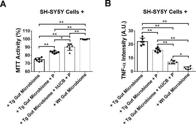
Fig. 5. Probing the gut-brain axis via cell culture mechanistic paradigm.
MTT activity assay revealed cultured SH-SY5Y cells under Wt gut homogenates displayed robust cell viability (A) with only trace levels of TNF-α (B). In contrast, exposure of SH-SY5Y cells to Tg gut reduced cell viability by about 25% and increased by about 10-fold TNF-α levels compared to Wt-exposed SH-SY5Y cells (***p < 0.05, ****p < 0.01). Tg gut+P or Tg gut+hUCB+P still also showed lower cell viability and higher TNF-α expression compared to Wt gut-exposed SH-SY5Y cells (***p < 0.05, ****p < 0.01), but they increased cell viability by about 13–20% and decreased TNF-α by about 27–68% compared to Tg gut-exposed SH-SY5Y cells (***p < 0.05, ****p < 0.01), with the Tg gut+hUCB+P more effectively sequestering neurodegeneration and inflammation than Tg gut+P (***p < 0.05, ****p < 0.01).

