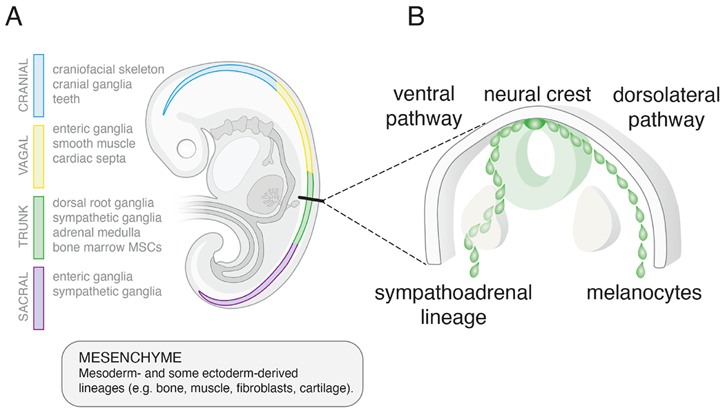
Figure 1. Extrinsic cues regulate neural crest lineage specification.

A) Drawing of a human embryo showing the neural crest (4-color). The cellular lineages derived from the neural crest vary based on their rostral-caudal location as shown (cranial (blue), vagal (yellow), trunk (green), sacral (purple)). Transplantation studies have shown that lineage specification is regulated primarily by the extrinsic cues rather than cell-autonomous factors. Trunk neural crest cells transplanted into the cranial region will produce cranial lineages and vice versa. Neural crest cells are multipotent and can give rise to both neuronal and non-neuronal (mesenchymal) cell types. This is particularly relevant for neuroblastomas that arise from the sympathoadrenal lineage of the trunk neural crest because they have both neuronal (N-type) and mesenchymal (S-type) cell populations. B) Drawing of a cross section of the trunk neural crest showing the two paths of migration. Both paths occur bilaterally but for simplicity the ventral pathway is shown on the left side of the embryo and the dorsolateral pathway is shown on the right. Extrinsic cues in these different regions of the embryo contribute to specification of the sympathoadrenal lineage and melanocytes. Thus, not only is the position along the rostral caudal axis important for neural crest derived cell lineage specification but the path of migration (ventral versus dorsolateral) is also important.
