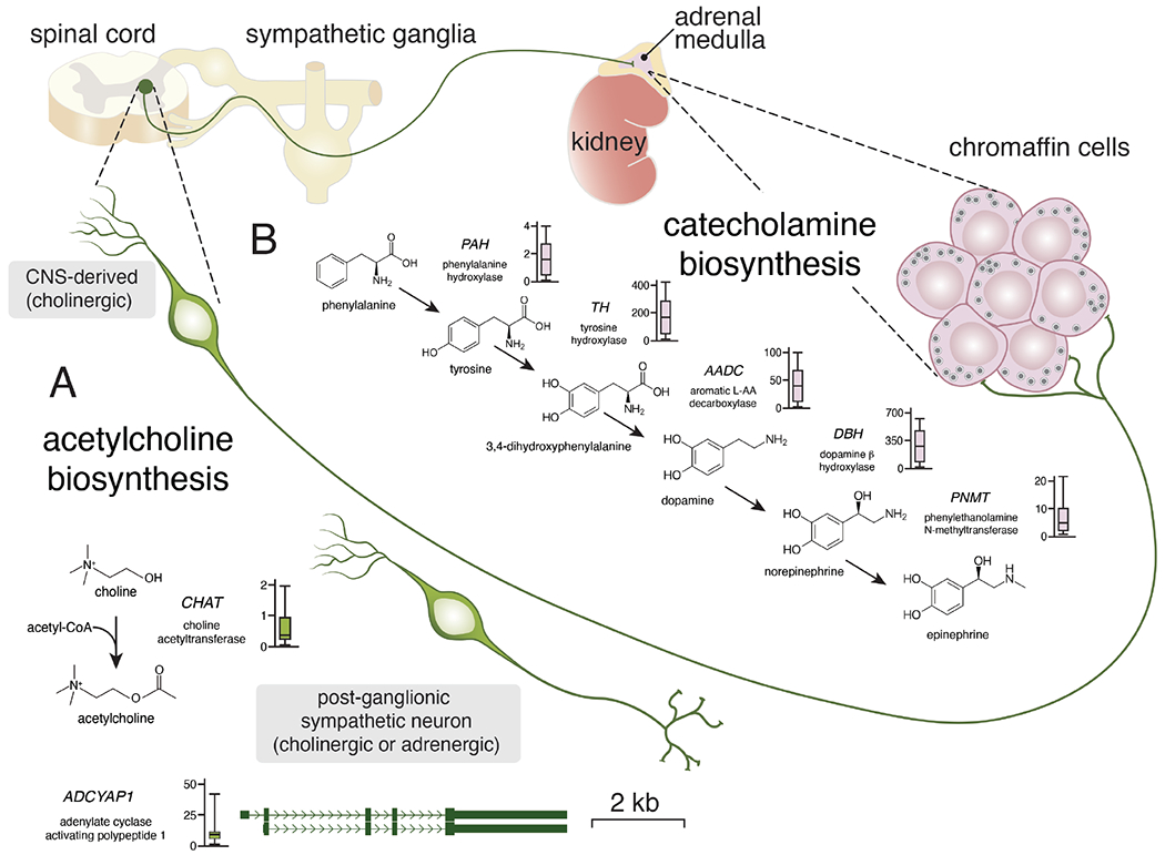
Figure 3. Expression of sympathoadrenal neurotransmitters and neuropeptides in neuroblastoma.

Simplified drawing of the neuronal connections in the sympathoadrenal lineage. Central nervous system (CNS) derived pre-ganglionic neurons synapse directly with chromaffin cells of the adrenal medulla where they release acetylcholine. Once stimulated, the chromaffin cells release epinephrine or norepinephrine into the circulation. Some chromaffin cells release epinephrine and some release norepinephrine from their dense core vesicles. A) Acetylcholine biosynthesis is regulated by choline acetyltransferase and the expression is relatively low (box plot) in the 191 representative neuroblastoma tumors. Acetylcholine is used for acute stimulation of the chromaffin cells. The neuropeptide encoded by ADCYAP1 is used for more chronic stimulation. While expression of ADCYAP1 is low in most neuroblastomas, a subset has very high levels of expression suggesting inter-tumor heterogeneity.
B) Drawing of the biosynthetic pathway for catecholamines with relevant gene and abbreviation for each enzyme. Next to each gene name is a boxplot of RNA expression (FPKM) from 191 neuroblastomas tumors (www.stjude.org/pecan). The chromaffin cells of the adrenal medulla secrete catecholamines as do a subset of post-ganglionic sympathetic neurons that are derived from sympathoblasts.
