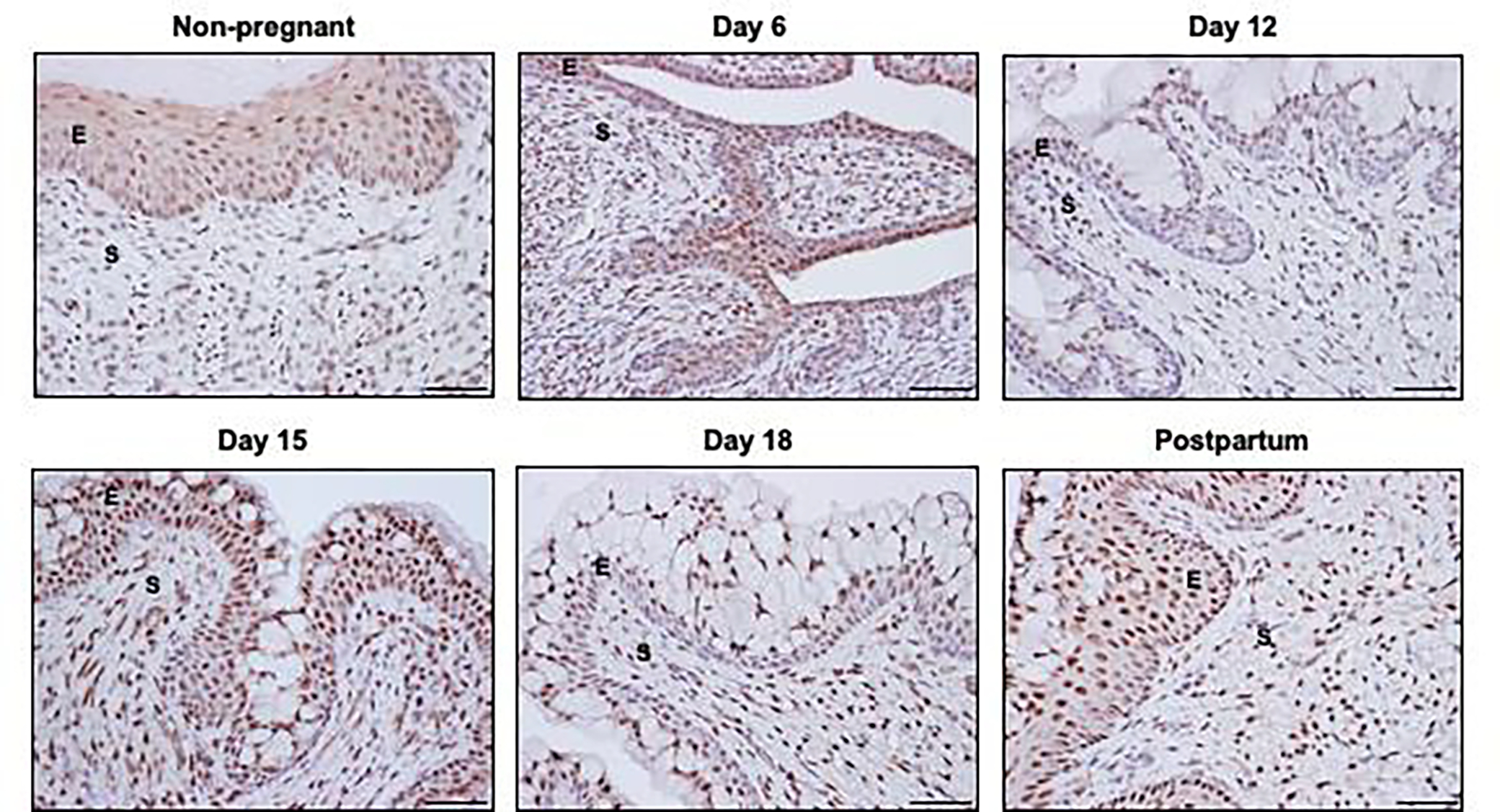
Figure 1: Progesterone receptor (PR) localization in non-pregnant and pregnant cervix.

Immunohistochemical localization of PR in cervical sections of non-pregnant (NP), pregnant and postpartum (PP) mice. Epithelial PR is localized throughout the basal and luminal layers of squamous epithelia in the NP and PP cervix. Similar patterns are noted in cervical sections from gestation days 6 and 15 while basal layers have reduced PR expression relative to the luminal secretory cells on days 12 and 18. Stromal PR remains high throughout pregnancy and postpartum compared to non-pregnant cervix. Images are taken at 20X. E= Epithelia; S= Stroma.
