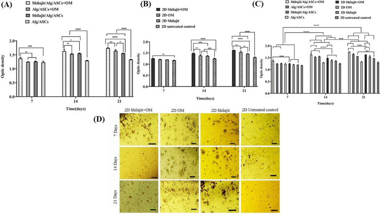
Fig. 6.
Detection calcium deposition. The calcium deposition assessment was done on 7, 14 and 21 days of differentiation in 3D (A) and 2D (B) groups and 3D conditions compared with matched 2D cultures in these time points (C). The highest level of calcium depositions was seen on Shilajit/Alg/ASCs + OM on 21 days after differentiation compared with other groups. D Micrographs of cells stained with alizarin red demonstrate the effects of shilajit on mineral deposition and nodule formation in 2D groups on 7, 14 and 21 days after differentiation. Qualitative observations showed that the number and size of the nodules increased in the presence of Shilajit (*P < 0.05, **P < 0.01, ***P < 0.001, ****P < 0.0001)

