Fig. 3.
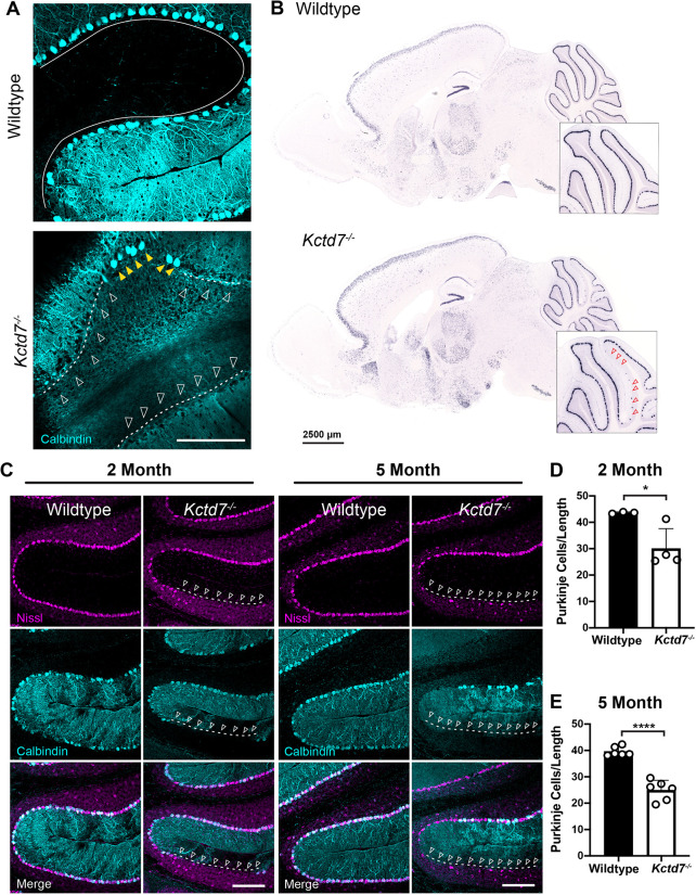
Kctd7 is required for Purkinje neuron survival. (A,B) The numbers and localization of calbindin-positive Purkinje neurons were assayed by immunohistochemistry analysis (A) and in situ hybridization (B) for calbindin in adult 2-month-old mice. In wild-type animals, Purkinje neurons form a single, continuous layer that clearly traces each cerebellar lobule (solid line). In Kctd7−/− mice, clear loss of Purkinje neurons is apparent in both visualization methods, as indicated by large gaps in the calbindin-positive layer [indicated by dashed lines and unfilled white (A) and red (B) arrowheads]. Yellow arrowheads in A indicate remaining Purkinje neurons in the Kctd7 mutant. (C-E) The distribution (C) and number (D,E) of Purkinje neurons were quantified at 2 and 5 months of age in wild-type controls (n=3 and 6 animals for 2 and 5 months, respectively) and mutant animals (n=4 and 6 animals for 2 and 5 months, respectively). Purkinje neuron numbers were significantly reduced at both time points, indicating that Purkinje neuron loss is an early feature in Kctd7−/− mice. Data are represented as the mean±s.e.m. *P<0.05; ****P<0.0001; unpaired two-tailed t-test for significance. Scale bars: 200 μm (A,C); 2.5 mm (B).

