Fig. 4.

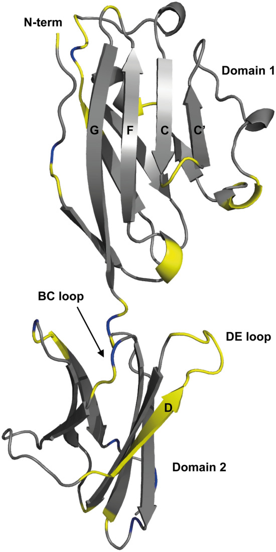
A ribbon representation of the backbone topology of PD-L1 (D1D2) (PDB: 3FN3), with the positions of residues with non-assigned backbone amide groups highlighted in yellow. The locations of proline residues are shown in blue

A ribbon representation of the backbone topology of PD-L1 (D1D2) (PDB: 3FN3), with the positions of residues with non-assigned backbone amide groups highlighted in yellow. The locations of proline residues are shown in blue