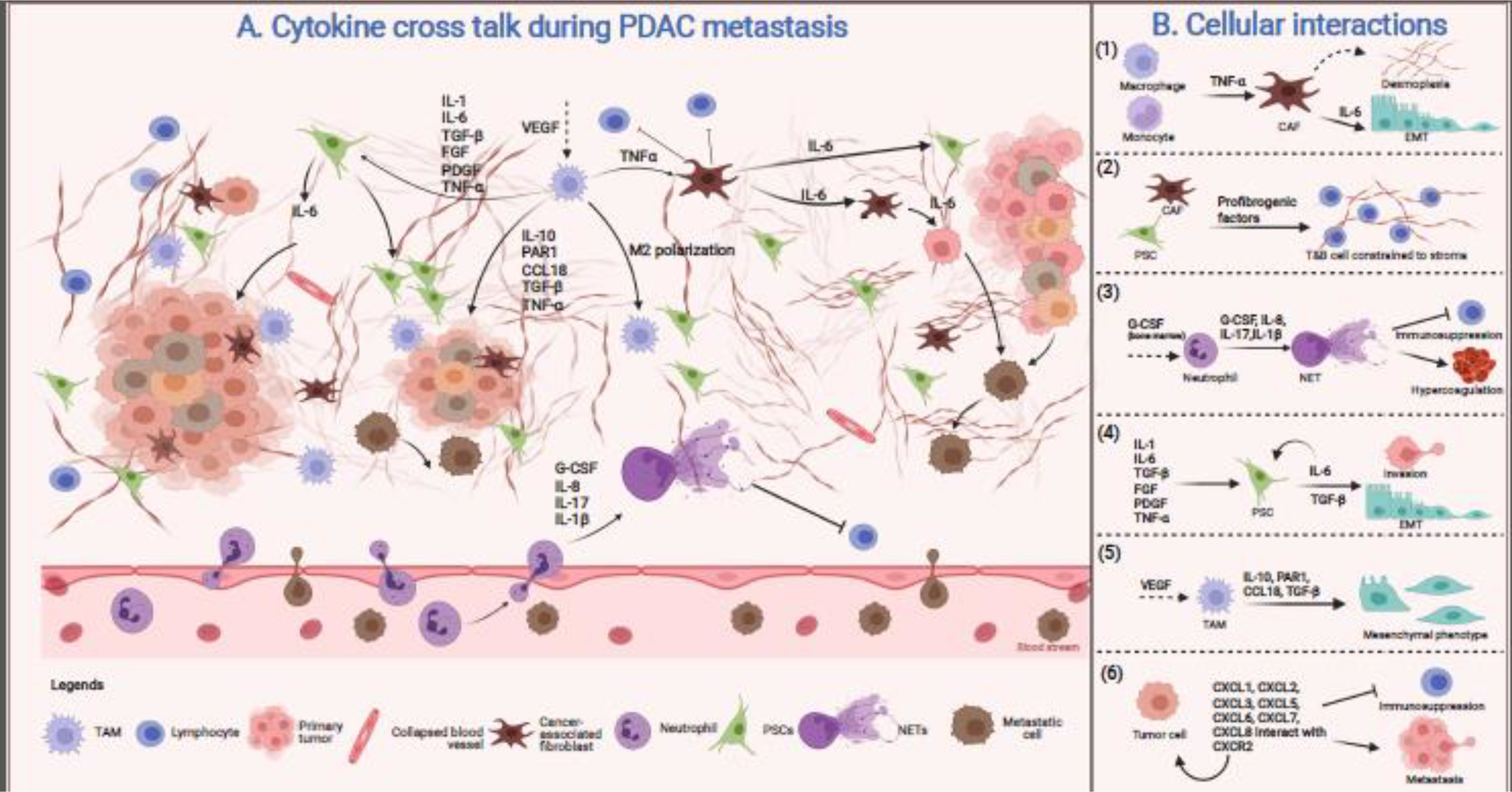
Figure 2: Cytokine crosstalk in PDAC tumor microenvironment.

A) The metastasis-promoting microenvironment is created by crosstalk between a labyrinth of versatile cells present in the TME. This complex network secretes various cytokines resulting in immune infiltration to facilitate migration and invasion. B) TNFα is an inflammatory cytokine secreted by macrophages and monocytes, which stimulates CAFs to secrete more fibrous stroma and participate in various signaling pathways promoting EMT (1). Cells such as CAFs and PSCs secrete profibrogenic factors that increase ECM deposition constraining lymphoid (B- and T-) cells to the tumor periphery, physically preventing their access to cancer cells (2). Secretion of G-CSF and other factors from the TME mobilize neutrophils from the bone marrow and triggers NET formation in the primary tumor and circulation. The NETs further promote immunosuppression as well as hypercoagulation (3). Cytokine milieu triggers PSCs to aid in invasion and EMT through an IL-6/STAT3 mediated pathway (4). The M2 polarized macrophages secrete inflammatory cytokines altering cancer cell morphology to a mesenchymal phenotype (5). Tumor cells secrete various chemokine, CXC-ligands, and express CXC-receptors, facilitating immunosuppression and dislodging of the cancer cells into the circulation for distant colonization (6). Figure created with BioRender.com.
