Figure 3.
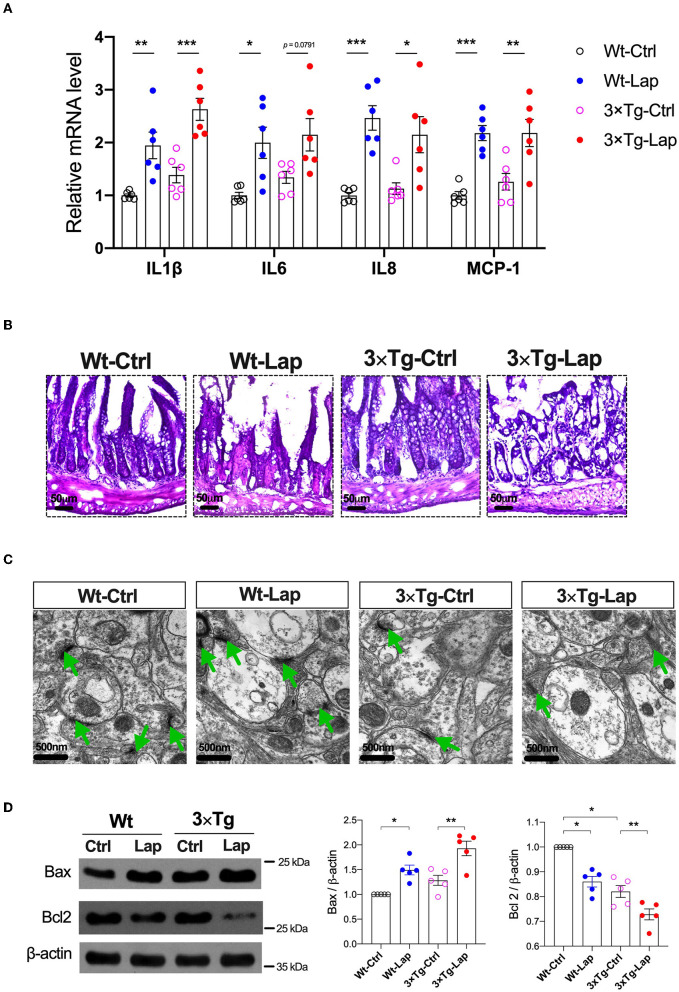
Laparotomy caused similar inflammatory responses in the periphery and hippocampus in both wild-type and 3×Tg AD mice. (A) Relative mRNA level of pro-inflammatory cytokines at postoperative 24 h in the hippocampi. IL-1β: F(3, 20) = 15.60, P < 0.0001; IL-6: F(3, 20) = 5.97, P = 0.0044; IL-8: F(3, 20) = 11.57, P = 0.0001; MCP-1: F(3, 20) = 13.18, P < 0.0001. (B) Morphological changes of acini and base of the intestine in postoperative 14 days by using H&E staining. Representative pictures were obtained from three independent experiments. (C) Representative ultrastructure of the hippocampal neuronal synapse using transmission electron microscopy following laparotomy in both wild-type and 3×Tg AD mice. (D) Protein expressions of apoptotic proteins, Bax, and Bcl2 in the hippocampi were determined by Western blot analysis. Bax: F(3, 16) = 14.79, P < 0.0001; BCL2: F(3, 16) = 33.36, P < 0.0001. Wt, wild-type mice; 3×Tg, triple transgenic mice; Ctrl, control; Lap, laparotomy. Data are presented as mean ± SEM and were analyzed using one-way ANOVA using Tukey's post hoc test, *P < 0.05, **P < 0.01, ***P < 0.001.

