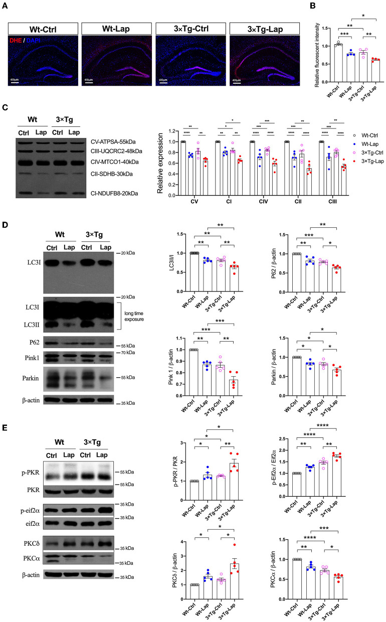
Figure 5.
Laparotomy impaired mitochondria mediated by the activation of PKR. (A) To evaluate the oxidative stress in the CNS, DHE staining was used in the entire hippocampal region following laparotomy in two strains of animals. Representative pictures were obtained from three independent experiments. (B) Mitochondria membrane potentiation was determined by JC-1 assay in the isolated mitochondria fractions in the hippocampi. F(3, 12) = 26.14, P < 0.0001. (C) After laparotomy, the expression of the mitochondria OXPHOS complex was detected by Western blot analysis in the hippocampi. CV:F(3, 16) = 27.28, P < 0.0001; CI: F(3, 16) = 35.95, P < 0.0001; CIV:F(3, 16) = 24.85, P < 0.0001; CII:F(3, 16) = 20.70, P < 0.0001; CIII:F(3, 16) = 21.64, P < 0.0001. (D) Western blots of autophagy-related proteins, including LC3, P62, Pink1, and Parkin, in the hippocampus following laparotomy in the middle-aged wild-type and 3×Tg AD mice. (E) Activities of PKR and PKC in the hippocampus induced by laparotomy in both wild-type and 3×Tg AD mice. Wt, wild-type mice; 3×Tg, triple transgenic mice; Ctrl, control; Lap, laparotomy. Data are presented as mean ± SEM and were analyzed using one-way ANOVA using Tukey's post hoc test, *P < 0.05, **P < 0.01, ***P < 0.001, ****P < 0.0001.

