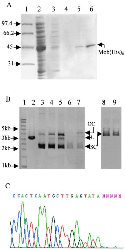
FIG. 5.
In vitro cleavage of supercoiled DNA by the purified Mob protein. (A) The Mob protein was tagged with six histidines at its carboxy-terminal end and purified. The proteins were separated in an SDS–10% polyacrylamide gel. Lanes: 1, protein standards (molecular sizes are given in kilodaltons); 2, 1 μl of crude extract of 15 ml of induced BL21(DE3, pLys, pETMobhis); 3, 25 μl of supernatant after the first wash of the His-resin; 4, 25 μl of supernatant after the second wash of the His-resin; 5, elution with 250 μl of buffer containing 50 mM imidazole (7.5 μl on the gel); 6, elution with 250 μl of buffer containing 125 mM imidazole (7.5 μl on the gel). (B) In vitro cleavage of supercoiled DNA. Plasmid DNA profiles after electrophoresis in a 0.9% agarose gel are shown. Lanes: 1, DNA molecular size marker (with sizes given in kilobases on the left); 2, restriction by PstI of the p2oriT plasmid DNA containing two oriTs in opposite directions (the linear plasmid has a size of 3,143 bp); 3, supercoiled p2oriT DNA (1 μg); 4, incubation of 1 μg of p2oriT DNA with the purified Mob protein (≅200 ng); 5, incubation of 1 μg of p2oriT DNA with the purified Mob protein (≅400 ng); 6, supercoiled poriT52bp DNA containing one oriT (0.5 μg); 7, incubation of 0.5 μg of poriT52bp DNA with Mob(His6) (≅200 ng); 8, supercoiled DNA of the control plasmid which contains no oriT (0.8 μg); 9, incubation of 0.8 μg of the control plasmid DNA with the purified Mob protein (≅200 ng). Note the increase of relaxed DNA (OC, open circle) in lanes 4, 5, and 7 and the partial linearization of p2oriT due to the cleavage of both oriTs in lane 5 (L, linear DNA; SC, supercoiled DNA). (C) Position of the nick site. Shown is an electropherogram of the sequence of the relaxed p2oriT DNA purified from the agarose gel using an automatic sequencer.

