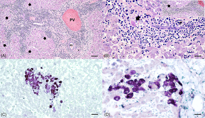
FIGURE 3.

Histopathological features of DCH‐positive liver sections of HPD cats. (A) The portal tracts were surrounded by thick bands of fibrosis that bridged between portal structures in some areas and contained high numbers of infiltrates of numerous lymphocytes and plasma cells. Infiltrates of lymphocytes and plasma cells extended beyond and disrupted the limiting plate (arrows). (B) Infiltrated lymphocytes and plasma cells clustered within the hepatic sinusoid, extended beyond and disrupted the limiting plate (inset), and dissected around adjacent hepatocytes, accompanied by rare acidophilic body (arrows) that represent interface hepatitis. Variable sizes of vacuolated hepatocytes were also presented. (C) Hybridization signals of DCH were present within most of the cytoplasm of hepatocytes in this field. (D) A cluster of DCH‐positive cells was present within the area of inflammation adjacent to the portal tract. An asterisk indicates hepatic artery. BD, bile duct; PV, portal vein. Bars indicates 45 μm for (A), 170 μm for (B, D), and 80 μm for (C)
