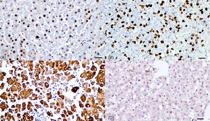
FIGURE 4.

Proliferative and apoptotic features of DCH‐positive liver section of HPD cats. (A) The PCNA and phospho‐histone H3 (B) immunostainings were diffusely presented within the nucleus of hepatocytes (golden brown precipitates), indicating proliferative activity of DCH‐infected liver. (C) Cytoplasmic immunostaining of survivin was positive within the cytoplasm of hepatocytes. (D) No immunostaining of cleaved caspase‐3 was found in the DCH‐positive liver section (red precipitates). Bars indicate 170 μm
