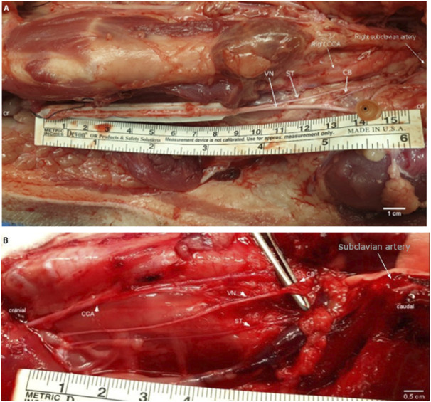
FIGURE 2.

(A). Surgical window of the right cervical and cardiac region in a pig. White arrows point at the VN, the ST, and the superior CB. The positions of the CCA and right subclavian artery are labeled for anatomical orientation. The inferior cardiac branch was not used in this study and is located caudal to the subclavian artery. Cr, cranial; cd, caudal; CCA, common carotid artery; VN, cervical vagus nerve; ST, sympathetic trunk; CB, superior cardiac branch. Cr, cranial; cd, caudal; CCA, common carotid artery; VN, cervical vagus nerve; ST, sympathetic trunk; CB, superior cardiac branch. (B). Surgical window of the right cervical and cardiac region in a rabbit. The cervical VN gives one superior CB to the heart. White arrows point at the VN; the ST; and the superior CB. Single white arrow pointing at position of the subclavian artery (behind rib cage). The inferior cardiac branch was not used in this study and is located below the subclavian artery. VN, vagus nerve; ST, sympathetic trunk; CB, superior cardiac branch; CCA, common carotid artery.
