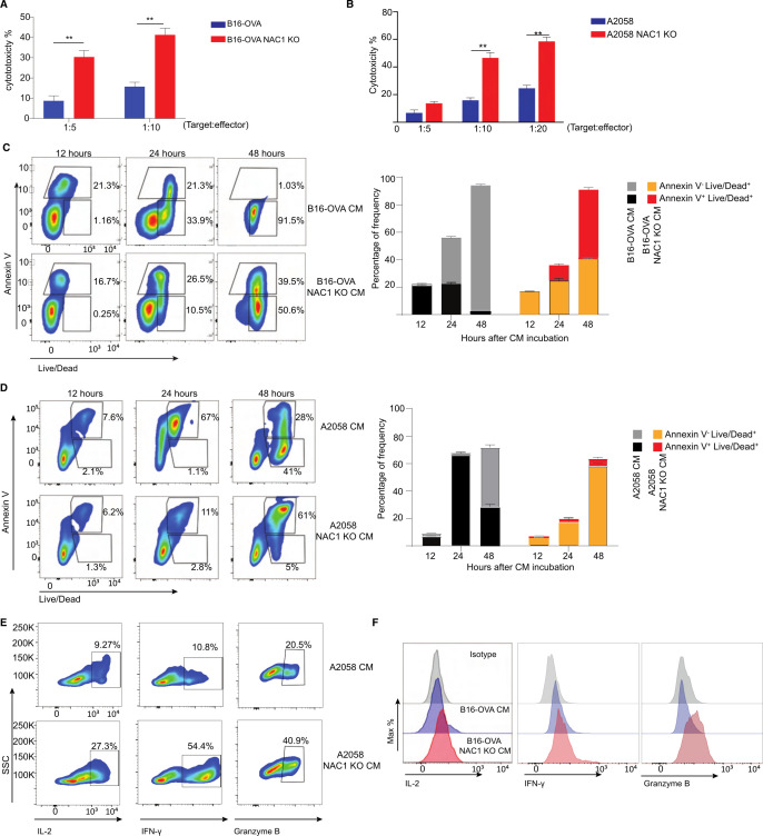
Figure 3.
Depleting tumorous NAC1 strengthens the cytotoxicity of CD8+ T cells. (A) WT or NAC1 KO B16-OVA cells (targets) were co-cultured with OT-I CD8+ T cells (effectors) (ratio: 1:5 and 1:10) for 6 hours. Per cent cytotoxicity was calculated as (1- (R5/R0)) x 100, where R5 = (target cells as % of total of 6 hours) / (effector cells as % of total of 6 hours), R0 = (target cells as % of total of 0 h) / (effector cells as % of total at 0 h). (B) WT or NAC1 KO A2058 cells were co-cultured with anti-tyrosinase TCR transduced human CD8+ T cells (effectors) (ratio: 1:5, 1:10, 1:20) for 6 hours. Per cent cytotoxicity was calculated as (1- (R5/R0)) x 100, where R5 = (target cells as % of total at 6 hours) / (effector cells as % of total of 6 hours), R0 = (target cells as % of total of 0 hour) / (effector cells as % of total of 0 hour). (C) Representative contour plots of Annexin V and Live-Dead expression on the CM-treated OT-I CD8+ T cells (left panel). Frequencies of the indicated Annexin V+ and Live-Dead+ expressing populations (right panel). After incubation with WT conditional medium (CM) or NAC1 KO B16-OVA CM for 12, 24, and 48 hours, respectively. Apoptotic rates were assessed by flow cytometry. One representative experiment out of three is shown. (D) Representative contour plots of Annexin V and Live-Dead expression on the CM-treated human CD8+ T cells (left panel). Frequencies of the indicated Annexin V+ and Live-Dead+ expressing populations (right panel). After incubation with WT CM or NAC1 KO A2058 CM for 12, 24, and 48 hours, respectively. Apoptotic rates were assessed by flow cytometry. One representative experiment out of three is shown. (E) Histograms showing expression of the indicated cytokines after incubation with WT CM or NAC1 KO B16-OVA CM for 6 hours (E); WT CM and NAC1 KO A2058 CM (F). One representative of three identical experiments is shown. IFN-γ, Granzyme B, and IL-2 expression were determined by flow cytometry. **p≤ 0.01; one way analysis of variance with multiple comparision correction. NAC1, nucleus accumbens-associated protein-1; OVA, ovalbumin.

