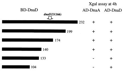
FIG. 2.
Interactions of various DnaD deletion proteins with DnaA and wild-type DnaD. Thick bars indicate DnaD portions fused to the Gal4 BD. These dnaD parts were amplified by PCR and cloned in pGBT9 as described for construction of BD-DnaD in the text. The carboxyl termini of the BD-DnaD proteins are shown by the amino acid positions in the wild-type DnaD at the right of the bars. Interaction was judged by change of color (+, blue, −, white) after 4 h of incubation in 5-bromo-4-chloro-3-indolyl-β-d-galactopyranoside (X-Gal). The location of the dnaD23 mutation is shown by the amino acid position (166th) with an arrow.

