
Figure. 1.
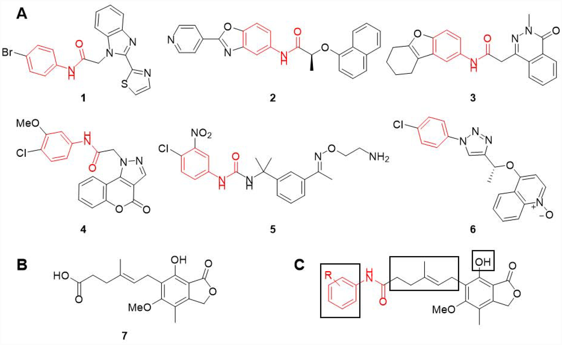
(A) Six structurally distinct CpIMPDH inhibitors (CpIMPDH IC50 = 12 – 64 nM).27 The fragments found in these inhibitors that interact with the adenosine subsite (A-site) of the NAD binding site are highlighted in red. This is based on co-crystal structures of 1, 2, 4 and 5 with CpIMPDH and 6 with Clostridium perfringens IMPDH (ClpIMPDH). The interactions of inhibitor 3 are assumed based on structural similarity since it has not been co-crystalized with an IMPDH. (B) Structure of mycophenolic acid (MPA, 7). (C) Mycophenolic anilides with three regions explored herein shown in boxes.