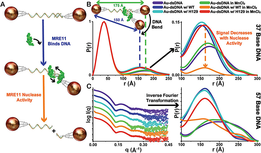
Fig. 5.
Demonstration of overall XSI assay scheme. (a) The proposed mechanism of MRE11 interaction with intact Au-dsDNA substrates and the subsequent nuclease activity leading to separation of the fixed inter-particle distances as Au-ssDNA. (b) Demonstration of the shifts in the distribution of inter-Au electron-pair distances, seen in the normalized P(r) functions (37-bp DNA), to lower mean values compared to the substrate alone representing the structural changes in the Au-dsDNA substrates associated with MRE11 binding. Additionally, a decrease in the amplitude of P(r) corresponding only in the inter-Au regions is observed only with WT-MRE11 in the presence of MnCl2 (orange) suggesting increased MRE11 nuclease activity (Au-dsDNA to Au-ssDNA). Legend for sample identity shown. (c) Exemplary experimental XSI curves and derived P(r) functions for 57-bp DNA colored as in the Panel (b). Curves have scaled I(q) for visualization purposes. P(r) function plot is scaled to show inter-Au distance region. All P(r) functions are normalized to the intra-Au peak to compensate for fluctuations in concentration as depicted in Fig. 1c

