Figure 1.

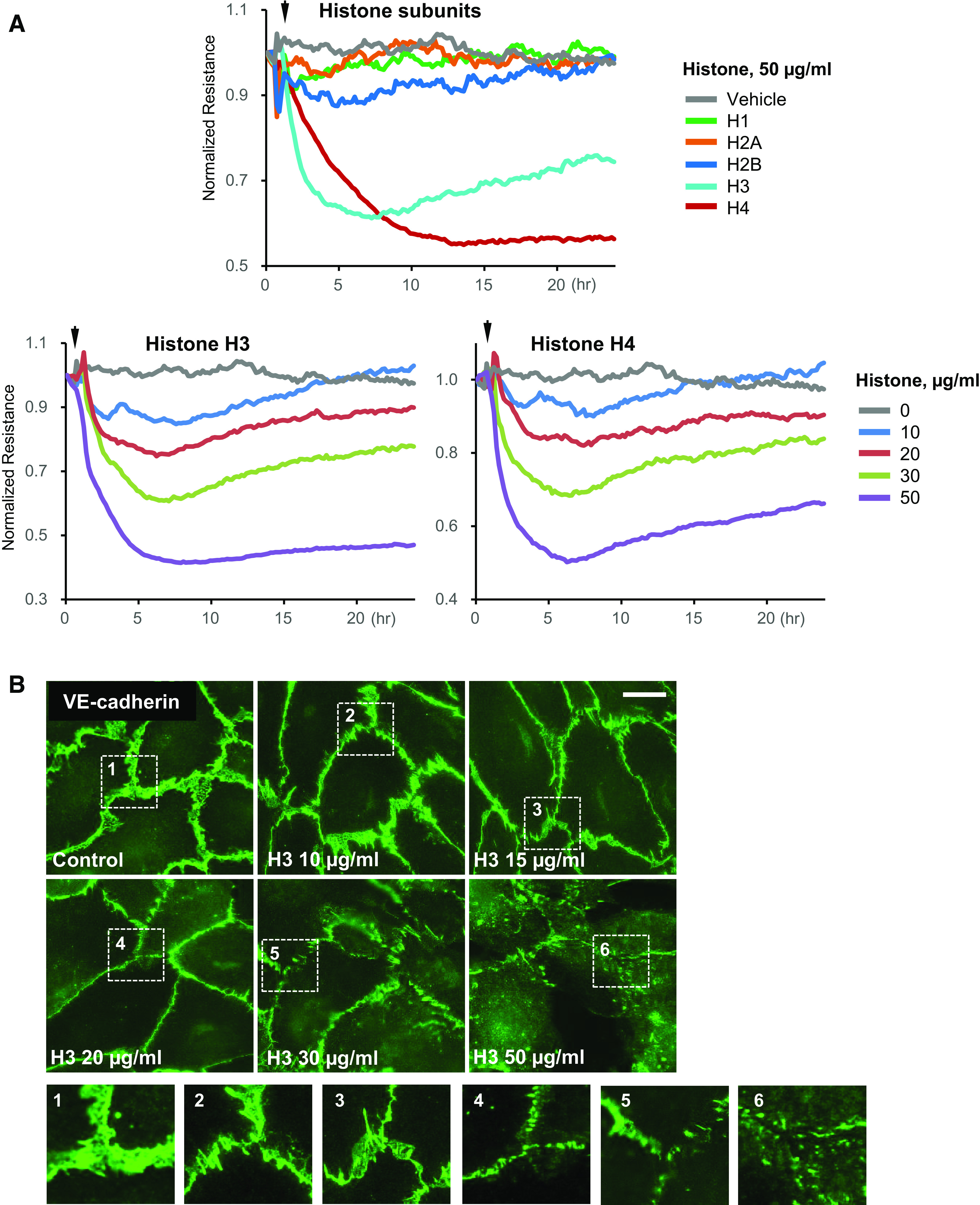
Selectivity of histone subunits in causing endothelial dysfunction. A: HPAECs were stimulated with indicated histone subunits, and endothelial permeability was determined by monitoring TER over time (top). The dose dependence of H3 and H4 on endothelial barrier disruption is also presented (bottom). Representative TER traces are shown for n = 8–10. B: HPAECs were treated with indicated concentrations of histone H3 for 4 h, and VE-cadherin immunostaining was performed. Insets: higher magnification images from the marked areas to illustrate the loss of VE-cadherin from cell membrane. Shown are representative images captured from 10 microscopic fields/condition (n = 3); bar = 5 µm. HPAECs, human pulmonary artery endothelial cells; TER, transendothelial electrical resistance.
