
Figure 4. DCs, but not activated TCD4+, indirectly present HIV-1-derived epitope.
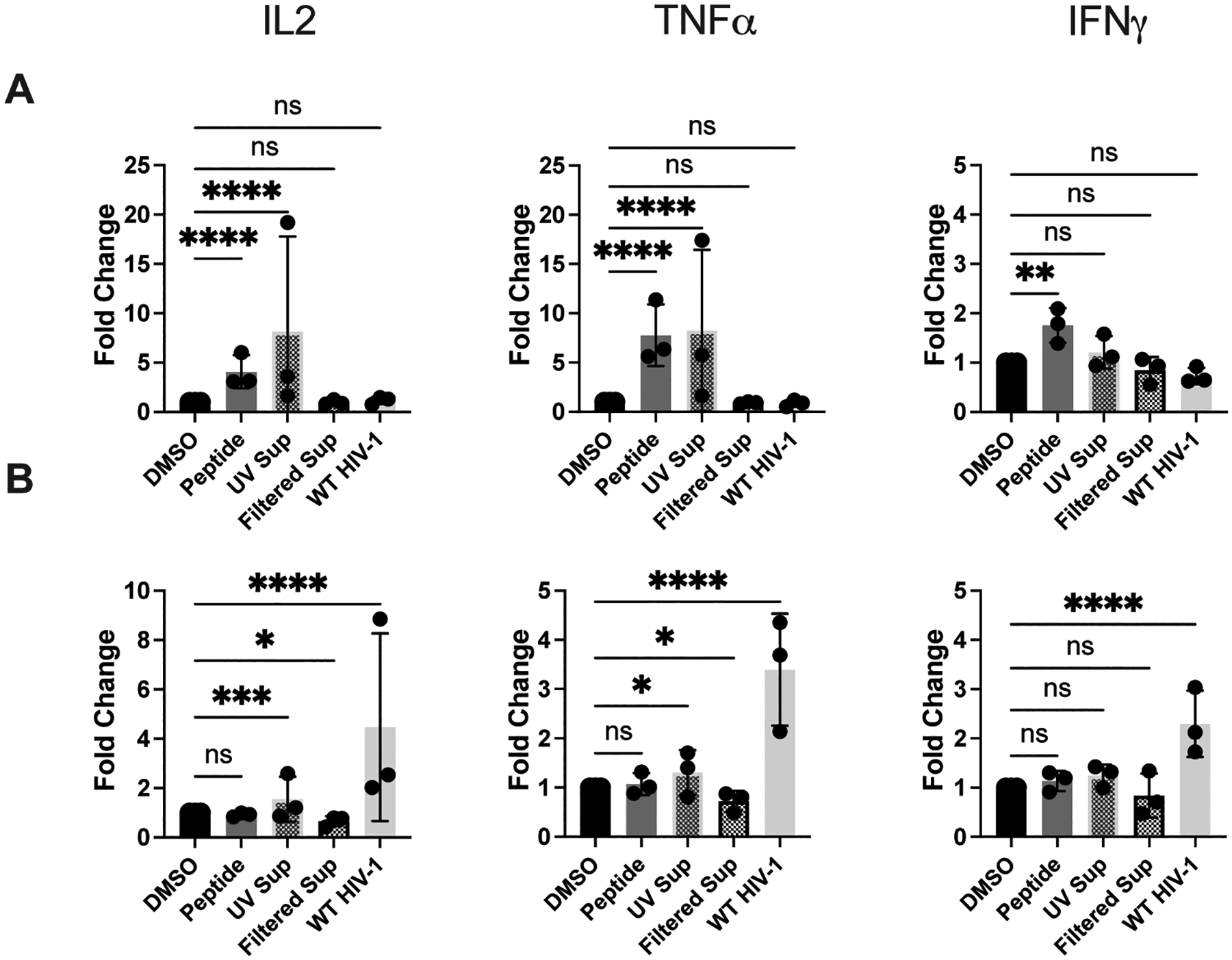
Presentation of UV-treated and filtered HIV-1+ supernatant by DCs and activated TCD4+ to HIV-1-specific TCD4+. DCs and activated TCD4+ were cultured and infected with WT HIV-1 as described in Materials and Methods. Supernatant from WT HIV-1-infected TCD4+ was collected and treated with UV light or filtered through a 100 kDa filter and then added to DCs and activated TCD4+ 12 hours prior to the beginning of the assay. (A) DCs and (B) TCD4+ were cocultured with HIV-1-specific TCD4+ for 8 hours and IL-2, TNFα, and IFNγ expression was assessed by flow cytometry. Fold induction of each cytokine is shown, using DMSO as a baseline. Each dot represents an independent experiment. Bars represent mean ± SD. One way ANOVA, *p<0.05, **p<0.01, ***p<0.005, ****p<0.001.