



Abstract
Retinoic acid-inducible gene I (RIG-I)-like receptors (RLRs) are cytosolic RNA sensors critical for initiation of antiviral immunity. Activation of RLRs following RNA recognition leads to production of antiviral genes and interferons (IFNs) for induction of broad antiviral immunity. While the RLRs are ubiquitously expressed, much of our understanding of these molecules comes from their study in epithelial cells and fibroblasts. However, RLR activation is critical for induction of immune function and long-term protective immunity. Recent work has focused on the roles of RLRs in immune cells and their contribution to programming of effective immune responses. This new understanding of RLR function in immune cells and immune programming has led to the development of vaccines and therapeutics targeting the RLRs. This review covers recent advances in our understanding of RLR contribution to immune cell function during infection and the emerging RLR-targeting strategies for induction of immunity against cancer and viral infection.
Keywords: Innate immunity, RNA helicases, Inflammation, Adaptive immunity
Introduction
The recognition of pathogen-associated molecular patterns (PAMPs) by membrane-bound and cytosolic pattern recognition receptors (PRRs) results in the induction of type I and III interferon (IFN) responses. IFNs play a critical role in providing antiviral protection through paracrine and autocrine induction of hundreds of effector antiviral genes and are critical to the initiation of adaptive immune responses (1–3). IFN induction is critical not only for antiviral immunity, but also induction of anti-tumor immunity and protection against bacteria, fungal, and parasitic infection (4–6). Further, aberrant expression of IFN promotes autoimmune diseases such as systemic lupus erythematosus (SLE), inflammatory myositis, Aicardi-Goutières syndrome (AGS), rheumatoid arthritis and other rheumatic diseases (7–9). Multiple therapeutics targeted at limiting IFN responses have been clinically developed and are in clinical trials or approved for treatment of specific autoimmune disorders (7, 8). As such, recombinant IFN has been developed as an antiviral therapeutic. However, IFN therapies are frequently accompanied by severe side effects, limiting clinical application (10). Given the dual role of IFN in promoting antiviral immunity while exacerbating autoimmunity, understanding the functions of RLRs beyond production of IFN is important for harnessing this pathway for therapeutic benefit. Particularly unravelling the cell types and mechanisms required for RLR promotion of adaptive immune responses have important implications for human health and disease (11, 12).
Nucleic Acid Sensing Pathways
IFNs are induced by multiple pathogen sensing pathways including the retinoic acid-inducible gene I (RIG-I)-like receptors (RLRs) (the focus of this review), toll-like receptors (TLRs), nucleotide-binding and oligomerization domain (NOD)-like receptors (NLRs), and the cyclic GMP-AMP synthase (cGAS)/stimulator of interferon genes (STING) DNA sensing pathways. These are localized in distinct cellular compartments and recognize unique ligands to allow for effective immunity against a variety of pathogens. These pathways have been described in detail in other recent reviews (13–16). TLRs are type I transmembrane proteins expressed at the plasma membrane or in endosomes/lysosomes that broadly recognize membrane components and nucleic acids derived from pathogens leading to activation of transcription factors, such as nuclear factor kappa B (NF-κB) and activator protein 1 (AP-1), to induce pro-inflammatory and IFN responses (13, 17–19). NLRs generally detect components of peptidoglycan and nucleic acid PAMPs in the cytosol, leading to type I IFN as well as interleukin 1 (IL-1) release (20–24). The cGAS/STING pathway recognizes host and foreign DNA for activation of TANK-binding kinase 1 (TBK1) and interferon regulatory factor 3 (IRF3), inducing a robust type I IFN response (16, 25, 26).
RLR Pathway
The RLRs are cytoplasmic RNA sensors that can initiate IFN, antiviral, and pro-inflammatory responses (27, 28). The family of RLRs consist of RIG-I, melanoma differentiation-associated protein 5 (MDA5), and laboratory of genetics and physiology 2 (LGP2), encoded by DDX58, IFIH1, and DHX58, respectively (29). All of the RLRs contain a structurally conserved DExD/H box RNA helicase and carboxy-terminal domain (CTD) (30). In addition, both RIG-I and MDA5 (but not LGP2) contain two amino-terminal caspase activation and recruitment domains (CARDs)(27) (Figure 1a). Interaction of cognate RNA ligands with the DExD/H helicase causes a confirmational change to displace the CARDs, allowing for their oligomerization and interaction with mitochondrial antiviral signaling proteins (MAVS) CARD domains (27, 29–31). MAVS is embedded in the mitochondrial-associated membranes (MAMs) and in oxidative organelles, peroxisomes (27). Canonically, MAVS forms a scaffold at MAM with tumor necrosis factor (TNF) receptor-associated factor 2 (TRAF2), TNF receptor-associated factor 5 (TRAF5), and TRAF6 to activate inhibitor of nuclear kappa-B kinase (IKK) family and TBK1 (32). IKK family kinases and TBK1 phosphorylate NF-κB, IRF-3 and IRF-7, which allows for their translocation to the nucleus and transcription of IFN, antiviral genes, pro-apoptotic genes, and immune cell programming genes (27). LGP2 plays a dual role in propagating MDA5 signaling during the acute stage of infection, but limits signaling downstream of MAVS during resolution of infection (33–35) (Figure 1b).
Figure 1. RLR domains, polymorphisms, and signaling.

A) The RLRs RIG-I, MDA5, and LGP2 all contain a C-terminal repressor domain (CTD) and a helicase domain. RIG-I and MDA5 contain two caspase activation and recruitment domains (CARDs). The numbered dashes represent relative locations of single nucleotide polymorphisms (SNPs) present in coding regions of the RLRs with numbers corresponding to Table I. B) Upon virus infection, RIG-I and MDA5 sense cognate RNAs and undergo confirmational change to allow for interaction with and activation of MAVS at mitochondrial-associated membranes (MAM) for downstream activation of NF-kB and IRF3 leading to antiviral gene programming, IFN production, immune cell programming genes, and pro-apoptotic genes. The role of LGP2 is still under investigation, but during acute infection it stabilizes and propagates MDA5 signaling. However, during resolution of infection LGP2 dampens RLR signaling.
As RLRs recognize RNA, many studies have focused on delineating features that allow for RIG-I, MDA5, and LGP2 discrimination of self vs non-self RNA. RIG-I preferentially recognizes short RNAs containing 5’ triphosphate or diphosphate groups (36–41). MDA5 recognizes long RNA duplexes in high molecular weight RNAs (41, 42). LGP2 binds RNA with high affinity regardless of their length or the modifications in the 5’ end, but its direct role in independent signal activation vs regulation of other RLRs remains unclear (43). In addition, these helicases are under control of regulatory enzymes and interacting proteins that can further enhance RLR activation (44–46). Despite these conserved features, RIG-I and MDA5 have been described to recognize self RNA and contribute to antiviral immunity as well as autoimmunity (47–51), and several recent reviews have comprehensively covered known RNA ligands of the RLRs (27, 52, 53). Overall, these studies have led to an understanding that each RLR plays unique and distinct roles in antiviral control and potentially immune programming (27, 53, 54).
Virus recognition and induction of immunity in infected target cells (epithelial, fibroblasts, and others) leads to antiviral programming and cytokine/chemokine production that promotes antigen presenting cell (APC) maturation and migration for adaptive immune programming (55). However, RLRs are expressed within nearly all cell types, suggesting both extrinsic (e.g. other immune cell-, epithelial cell-, or fibroblast-mediated) and intrinsic roles for RLRs in immune cell function (27). While relatively few studies have focused specifically on RLR functions in specific immune cell subsets, there has been a renewed interest in delineating immune cell roles of these pathways with a focus on developing therapeutics and vaccine adjuvants that target host immune pathways normally activated following virus infection.
RLR Polymorphisms and Disease
Single nucleotide polymorphisms (SNPs) in RIG-I (DExD-Box Helicase 58, DDX58), MDA5 (Interferon Induced With Helicase C Domain 1, IFIH1), and LGP2 (DExH-Box Helicase 58, DHX58) have been identified with linkage to disease susceptibility (Figure 1a and Table I). For example, minor alleles of DDX58 (rs10813831 and rs3824456) and DHX58 (rs2074160) are associated with spontaneous clearance of hepatitis C virus (HCV) and hepatitis B virus (HBV) (56, 57). Conversely, minor alleles of DDX58 (rs3739674) and IFIH1 (rs1990760) are associated with severe enterovirus 71(EV71) infection or more severe HCV following solid organ transplant (58, 59). Despite their canonical role in antiviral immunity, SNPs in DDX58, IFIH1, and DHX58 are linked to enhanced risk of developing autoimmune diseases such as atypical singleton-merton syndrome, systemic lupus erythematosus (SLE), and type 1 diabetes (T1D) (60–65). However, several of these SNPs are protective against psoriasis/psoriatic arthritis and T1D, highlighting the critical role for appropriate RLR function in regulating inflammation and immunity (59, 66–68).
Table I.
RLR Polymorphisms and Disease Implications1
| # | SNP | Genotype | Mutation | Disease Implications | Source |
|---|---|---|---|---|---|
| RIG-I (DDX58) | |||||
| 1 | rs3739674 | G>C | Upstream mutation; reduced expression | Increased EV71 severity | (58) |
| 2 | rs10813831 | G>A | Coding mutation; missense variant | Spontaneous HCV Clearance; Conversely, Severe Recurrent HCV following Liver Transplant | (56, 59) |
| 3 | rs3824456 | C>G | Intron mutation | Spontaneous HBV Clearance | (57) |
| 4 | rs55789327 | G>A | Coding mutation; Missense variant | Rotavirus vaccine IgA seroconversion | (69) |
| 5 | rs786204848 | G>T | Coding mutation; missense variant | Atypical Singleton-Merten Syndrome; Congenital Glaucoma | (60) |
| 6 | rs786204847 | A>C | Coding mutation; missense variant | Atypical Singleton-Merten Syndrome | (60) |
| MDA5 (IFIH1) | |||||
| 7 | rs10930046 | T>C | Coding mutation; Missense variant | Decreased Psoriasis Risk; Increased SLE Risk | (59, 63, 66) |
| 8 | rs35744605 | C>A | Coding mutation; loss of function | Reduced T1D Risk | (67) |
| 9 | rs3747517 | T>C | Coding mutation; Missense variant | Increased SLE Risk | (61, 62) |
| 10 | rs35667974 | T>C | Coding mutation; Loss of function | Decreased Psoriasis and Psoriatic Arthritis Risk; Reduced T1D Risk | (66–68) |
| 11 | rs1990760 | C>T | Coding mutation; decreased expression, increased methylation | Increased T1D and SLE Risk; Increased EV71 Severity | (58, 61–64) |
| 12 | rs2111485 | A>G | Intergenic mutation; unknown, no expression change | Increased T1D Risk; Spontaneous HBV Clearance | (57, 64) |
| LGP2 (DHX58) | |||||
| 13 | rs12600570 | C>T | Intronic; unknown | Elevated Fasting Plasma Glucose | (65) |
| 14 | rs2074160 | G>A | Coding mutation; unknown | Spontaneous HBV Clearance | (57) |
While many associations with autoimmunity and chronic viral infection have been described, how RLR SNPs regulate susceptibility to these illnesses is not well understood. However, the emergence of novel mouse models containing SNPs equivalent to those found in humans will shed further light on the importance of these RLR SNPs in autoimmune and infectious disease and allow for greater understanding of their mechanism. For example, the IFIH1 rs1990760 minor allele is associated with multiple autoimmune diseases, but the mechanisms underlying this enhanced risk due to this polymorphism had not been elucidated. A murine model expressing risk (rs1990760 minor allele) and non-risk (rs1990760 major allele) alleles of IFIH1 was developed to determine the role of these haplotypes in IFN regulation, antiviral immunity, and susceptibility to autoimmune disease (62). Mice expressing the risk allele had higher basal IFNβ expression, enhanced protection against encephalomyocarditis virus (EMCV) infection, as well as increased autoantibody production and incidence of diabetes. Similar models and mechanistic studies will broaden our understanding of the implications of RLR SNPs in pathogen resistance and autoimmunity and allow for delineation of how these variants impact immune cell function.
For example, epistatic interaction between the rs55789327 minor allele of DDX58 and a polymorphism in TLR3 is associated with enhanced IgA seroconversion following rotavirus vaccination (69). However, the specific contribution of RIG-I to this process and in which cell types it is important remains unknown. Based on the relative number of peer-reviewed publications, much of our understanding of RLR regulation derives from in vitro studies on epithelial cells and fibroblasts. Thus, it is less well understood how cell identity further contributes to the functions and regulation of RLRs. As immune cells are critical effectors of both viral protection and drivers of autoimmunity, understanding the specific roles of RLRs in immune cells is critical for understanding the mechanisms of protective vs deleterious RLR responses and allow for design of targeted therapeutics promoting health while minimizing deleterious tissue damage. Recently, multiple studies have begun to unravel the roles of the RLRs and their functions in programming immune cell-specific responses, described below (Figure 2).
Figure 2. RLR functions in immune cell subsets.
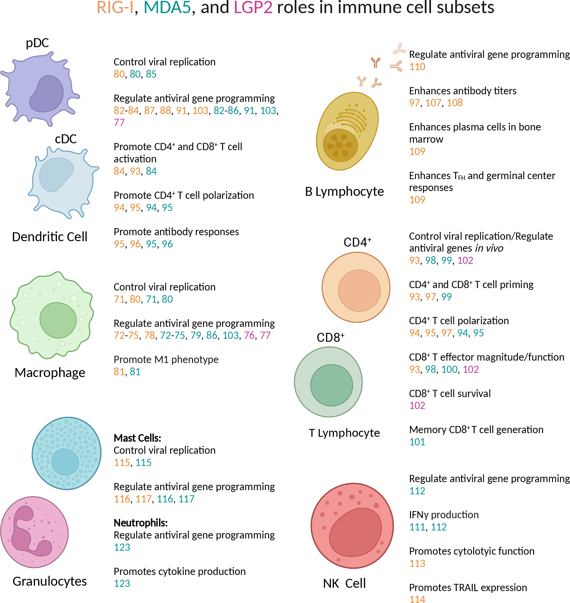
The roles of RIG-I, MDA5 and LGP2 in dendritic cells, macrophages, natural killer cells, B lymphocytes, T lymphocytes, and granulocytes during infection are summarized. References for each function are color-coded: RIG-I (orange), MDA5 (teal), LGP2 (magenta).
RLR Regulation of Immune Cell Function and Antiviral Responses
Macrophages
Macrophages play a vital role in controlling viral infection through the induction of downstream ISGs and pro-inflammatory cytokines. However, infected macrophages are disabled in their cytopathic function and require RLR sensing to control viral load (70). Murine bone marrow macrophages (BMMs) require MAVS downstream of RIG-I and MDA5 to induce type I IFN and pro-inflammatory cytokine expression in response to Vesicular Stomatitis Virus (VSV)(71). There is also evidence that both RIG-I and MDA5 contribute to production of MIP1α, RANTES, CXCL10 and IFNγ in human monocyte derived macrophages (MDM) during Middle East respiratory syndrome coronavirus (MERS-CoV) infection (72). Porcine alveolar macrophages depend upon both RIG-I and MDA5 in a redundant fashion to induce IFNβ and interferon stimulated gene 15 (ISG15) in response to African Swine Fever Virus (ASFV), VSV, Sendai Virus (SeV), EMCV, and Influenza A Virus (IAV), which suggest overlapping function between RIG-I and MDA5 in the context of these infections (73, 74). Further, in vivo murine model of Ebola virus infection found the RLR signaling adapter MAVS is required for secretion of IFNβ and protection against lethality (75). While the specific role of LGP2 in regulating RLR signaling is unclear, studies suggest LGP2 synergizes with RIG-I and MDA5 to promote proinflammatory cytokine and IFN induction in murine and porcine macrophages (76, 77) (Figure 1b).
Despite the canonical role for the RLRs in regulating antiviral immunity in macrophages, there is evidence that the RLRs play unique roles in regulating virus infection and macrophage function. RIG-I, but not MDA5, is required in human monocyte-derived macrophages for type I and type III IFN induction following H5N1 IAV infection (78). Murine BMM lacking MDA5 have diminished IFNβ production compared to wild-type and RIG-I deficient BMM during Murine Hepatitis Virus (MHV) infection (79). These differential requirements are due to the specific RNAs produced by these viruses during infection. Importantly, utilization of whole body murine genetic double knockouts of MDA5 and RIG-I demonstrate that both RLRs have non-redundant roles in mediating protection against West Nile Virus (WNV) as measured by replicating virus and diminished IFNβ production (80, 81). During WNV infection, the RLRs reinforce pro-inflammatory M1 macrophage programming, while suppressing wound healing M2 macrophages in both murine and human cells (81). Overall, these studies show that the RLRs regulate not only IFN and antiviral gene programming but play a direct role in regulating macrophage polarization.
Dendritic Cells
Among APCs, dendritic cells (DCs) play a critical role in presenting foreign antigen for the activation of adaptive immune responses important for pathogen clearance and long-term memory protection. While extrinsic inflammation induced by RLR signaling in epithelial and other cells impacts DC activation, there is evidence that DC activation, maturation, and antigen presentation capability are dependent upon intrinsic RLR signaling. Human monocyte derived DCs (moDCs) infected with IAV or Measles Virus rely on MDA5 and RIG-I for type I IFN induction, similar to murine bone marrow-derived DCs (BMDCs) during rabies virus and WNV infection (80, 82–84). MDA5 signaling in BMDCs is required for cytokine production during norovirus and EMCV infection (85, 86). LGP2 deficient DCs infected with reovirus, EMCV, SeV, and VSV have diminished production of type I IFN, thereby suggesting LGP2 regulates MDA5 and/or RIG-I signaling (77) (Figure 1b).
While human moDCs and murine BMDCs serve as tractable models, there is evidence that different DC subsets present in vivo rely differentially upon PRR pathways. Infection of murine embryonic fibroblasts (MEF) and conventional DCs (cDC) with Newcastle disease virus (NDV) led to a RIG-I-dependent induction of type I IFN. By contrast, the absence of RIG-I did not affect the production of IFN in plasmacytoid DCs (pDC). In pDC, NDV-induced IFN expression required the expression of the RNA sensors TLR7 and TLR9 (87). However, treatment of human pDC with a RIG-I activating HCV-derived PAMP led to production to type III IFN (88). Mass spectrometry analysis of splenic DC subsets revealed that RIG-I and MDA5 was readily detected in CD4+ and double negative (CD4−CD8α−) cDC subsets, but low or undetectable in CD8α+ cDC, suggesting that RLR expression unique to specific DC subsets may regulate their functions during acute viral infection (89). In addition, differences in the expression and utilization of signal transduction molecules downstream of PRRs across dendritic cell subsets could further dictate the cell type specific regulation of dsRNA detection (90).
Temporal changes in RLR expression may also allow for varying roles of RIG-I, MDA5 and/or LGP2 in specific cell subsets over the course of infection. During in vivo lymphocytic choriomeningitis virus (LCMV) infection, induction of type I IFN (day 1 post infection) is RIG-I-dependent, while the later IFN response (day 2 post infection) is MAVS-MDA5-dependent (91). Human DCs upregulate RIG-I, MDA5, and LGP2 following Zika Virus, suggesting these RLRs may contribute differentially to DC function and immune responses as infection progresses (92). Together, this supports that abundance of nucleic acid sensors, co-regulatory molecules, and downstream effectors all play a critical role in the spatial and temporal antiviral functions of RLRs. As we continue to uncover additional functions for RLRs in coordinating innate and adaptive responses beyond antiviral control, focusing on understanding their unique roles in immune cells will allow for the development of targeted therapeutics and adjuvants to fine-tune DC function for optimal activation of adaptive immunity.
In support of this, the RLRs play a role in regulation of DC function for downstream activation of adaptive immunity. RIG-I and MAVS signaling in DCs are required for generation of cytolytic CD8+ T cells function following IAV infection in vitro and in vivo (84, 93). Particularly, RIG-I signaling in murine BMDCs is necessary for presenting antigen to both CD4+ and CD8+ T cells during IAV infection (93). RLR signaling in dendritic cells also regulates CD4+ T cell immunity. Upon infection with dengue Virus, human DCs become activated to secrete IL-27, leading to polarization of TFH and production of IgM and IgG in a RIG-I and MDA5-dependent manner as shown by siRNA knockdown of MAVS (94, 95). Full MAVS knockout or deletion of MAVS in CD11c+ cells, but not B cells, led to increased virus titers and enhanced WNV-specific IgG and neutralizing antibody responses (96). Overall, RLR signaling in DCs is required not only for antiviral activity, but also support DC activation and generation of robust adaptive immune responses.
T cells
APCs, primarily DCs, play a critical role in regulating T cell activation. As such, both cell intrinsic and extrinsic dependencies on RLR expression have been described for T cell activation and function across various infectious models. RLRs can play non-redundant roles in CD4+ T cell activation. RIG-I is required for priming of CD4+ T cells during IAV infection in mice (93). During H5N1 IAV infection RIG-I is required for priming of CD4+ T cells and Th1 polarization (97). During dengue virus infection, both RIG-I and MDA5 are required for Th1 polarization (94). Interestingly, the absence of RLR expression in DCs appeared to be required for Th1 polarization during dengue virus. Further, both RIG-I and MDA5 in DCs are required for CD4+ TFH responses during dengue virus infection (95). TFH play an important role in germinal centers to support B cell and antibody-mediated responses. Therefore, it is likely this role for RIG-I and MDA5 in regulating TFH also impacts the development of B cell and antibody responses during infection.
While the RLRs play a role in CD4+ T cell function, much more is known about RLR regulation of CD8+ T cell responses. During IAV infection, RIG-I is important for CD8+ T cell priming, and mice lacking MAVS and RIG-I have reduced CD8+ T cell production of IFNγ, TNF, and Granzyme B (93). Mixed bone-marrow chimeras (lethal irradiation of wild type mice and reconstitution with RIG-I deficient bone marrow) and in vitro studies showed that RIG-I in both DCs and CD8+ T cells is required for CD8+ T cell priming and function during IAV infection (93). Mice lacking MDA5 have dysfunctional effector CD8+ T cell responses during WNV and MHV infection (98). During WNV infection, T cell extrinsic MDA5 signaling is required for CD8+ T cell priming to control CNS infection (99). Additionally, virus specific CD8+ T cell responses were significantly reduced in MDA5 deficient mice following chronic LCMV infection (100). This study showed MDA5-dependent induction of type I IFN extrinsic of T cells was required to prevent CD8+ T cell exhaustion (100). MDA5 also contributes to memory generation as MDA5 expression in non-hematopoietic cells enhanced survival and memory of CD8+ T cells during herpesvirus infection (101). LGP2 also contributes to regulation of T cell immunity. During WNV infection, WNV-specific CD8+ T cell populations and cell survival are reduced in mice lacking LGP2, but whether LGP2 functions intrinsically in CD8+ T cells or extrinsically in another cell type, such as an APC, to regulated WNV-specific CD8+ T cell immunity is currently unknown (102).
It is important to note that the individual contributions of RIG-I, MDA5, and LGP2 cannot be directly compared in vivo as commonly used mice deficient in RIG-I are embryonic lethal in a full C57BL/6 background and have to be maintained on a mixed Sv129 × C57BL/6 background or on an Sv129 background (87, 103, 104). As MDA5 and LGP2 deficient mice are on the C57BL/6 background, currently any comparison of these mice compared to those lacking RIG-I include the caveat that differences in experimental models may be due to background strain differences. Additionally, a lack of floxed RLR mice has prevented the investigation of the RLRs in individual cell subsets to date. However, regardless of whether the contribution is intrinsic to T cells or extrinsically regulated by RLR signaling in cells known to regulate T cell immunity, RLRs clearly contribute to T cell immunity. Given this, the RLRs have been targeted, particularly in the context of cancer, to prime cytotoxic anti-tumor T cell immunity (11, 12, 105, 106).
B cells
Our understanding of the mechanisms by which RLRs support B cell responses is starting to emerge, and multiple studies have demonstrated that adjuvant use of RLR agonists can promote B cell and antibody responses. Administration of RIG-I agonist RNA as an adjuvant administered with H5N1 virus-like particles to mice led to a decrease in viral titers and an increase in IgG2a responses (97). Inclusion of a RIG-I agonist in an influenza virus DNA vaccine vector led to activation of IFNβ in cell culture and increased serum antibody levels (107). The use of in vitro transcribed defective interfering RNA from SeV as an adjuvant for H1N1 IAV vaccination led to increased IgG subtypes, IgA in the airways, and enhancement of survival following lethal challenge (108). In addition to enhancement of antibody titers, one study utilizing administration of a RIG-I targeting adjuvant in combination with H1N1 IAV vaccination to mice found it led to a higher IgG antibody titer, enhanced germinal center formation, and earlier accumulation of virus-specific plasma cells in the bone marrow compared to vaccination alone (109). These results suggest targeting of RIG-I as an adjuvant may not only increase antibody titers, but also may enhance the durability of these responses. In humans, one polymorphism of RIG-I is correlated with enhanced IgA seroconversion following vaccination against rotavirus infection (69). Together, these studies indicate an important role for the RLRs in regulating B cell immunity that can be harnessed to boost protective immunity, but to date these studies have not delineated whether RIG-I signaling in DCs and/or B cells is required for this adjuvant effect. While it is likely RLR regulation of DC function contributes to this effect, B cells isolated from peripheral blood mononuclear cells of HCV positive patients displayed upregulation of both protein and mRNA transcripts for RIG-I and IRF-2, suggesting there may be an intrinsic role for RIG-I in regulating B cell responses during HCV infection (110).
Natural Killer Cells
There are few studies addressing the role of RLRs in natural killer (NK) cells during virus infection. However, the effects of synthetic agonists on viral sensors, such as RLRs, in NK cells has been investigated. Human and murine NK cells co-cultured with DCs and treated with poly(I:C), a synthetic dsRNA analogue known to activate MDA5, promotes IFNγ production by NK cells (111). This IFNγ production by NK cells is significantly reduced with the loss of MAVS and is dependent upon DCs (111). As poly(I:C) also activates the membrane-bound dsRNA sensor, TLR3, defining the target of this ligand that modulates NK function is necessary. Injection of mice with poly(I:C) led to IFNγ production in NK cells that was dependent upon both MDA5 and TLR3 (112). Mixed bone marrow chimera mice identified a requirement for MDA5 in the stromal compartment for NK cell type I IFN production in response to poly(I:C) in vivo (112). As observed in murine models, RLR agonists can also activate human NK cells (111). Human NK cells infected with HBV and treated with poly(I:C) have diminished levels of perforin, Granzyme B and IFNγ, compared to control (113). HBV infection reduced RIG-I expression and dampened NF-kB and mitogen activated protein kinase (MAPK) signaling (113). Targeting RIG-I activation by 5’triphosphate ssRNA treatment of NK cells led to increased surface expression of TNF-related apoptosis inducing ligand (TRAIL), suggesting there is also an NK cell intrinsic RLR role (114). Together these studies demonstrate a requirement for both RIG-I and MDA5 in regulating NK cell function.
Granulocytes
Mast cells primarily reside in the skin and mucosal tissues, and act as immune sentinels for various types of viruses. The expression of RLRs in mast cells is vital for initiating a state of inflammation to combat replicating virus. Silencing of MDA5 and RIG-I using small interfering RNA (siRNA) in human mast cells led to an increase in VSV titer during in vitro infection (115). Further, both RIG-I and MDA5 are required for murine mast cell production of IFNα, IFNβ, CXCL10, and IL-6 production during VSV infection (116). Additionally, knockdown of MDA5 and RIG-I sensors in KU812 mast cell line diminished CXCL10, CCL4, and CCL5 in response to antibody-enhanced dengue Virus (117). Primary human cord blood-derived mast cells infected with dengue virus in the presence of dengue immune sera led to an increase in mRNA expression of MDA5, RIG-I, as well as type I IFNs (117). Together, these studies support an important and potentially inducible role for RIG-I and MDA5 in regulating anti-viral responses and chemokine/cytokine production in mast cells.
Eosinophils contribute to fungal and allergic disease where they secrete chemokines/cytokines, degranulate, and form extracellular traps (118). However, whether eosinophils can respond to challenge for control of viral infection is an emerging area of research (119). Murine models have shown that RLR mRNA expression is lower than that observed in DCs (119). Expression of both Ddx58 and Ifih1 was induced by in vitro infection with IAV H1N1. Similarly, human eosinophils have low expression of RIG-I and MDA5 compared to expression of other PRRs known to promote eosinophil activation, such as NOD1 and NOD2 (120, 121). However, upon IAV challenge RIG-I mRNA expression can be rapidly induced, suggesting there may be a temporal role for RIG-I in eosinophils following upregulation of the receptor (122).
Neutrophils rapidly infiltrate infected tissue to phagocytose pathogens, debris, and cells and form extracellular traps to aid in clearance of viral infection. Transfection of human neutrophils with poly(I:C) to activate MDA5 results in upregulation of type I IFN, TNF, IL-12, multiple chemokines/cytokines, and ISGs, demonstrating neutrophils sense and respond to virus infection in a canonical fashion (123). IFNβ and TNF production by murine neutrophils during EMCV infection is dependent upon MDA5 (123). Human neutrophils treated with single-stranded RNA (ssRNA40) derived from Herpes Immunodeficiency Virus-1 (HIV-1) led to upregulation RIG-I and MDA5, similar to elevated levels in neutrophils isolated from HIV-1 positive patients (124, 125). Intriguingly, RIG-I and MDA5 have been observed in secretory vesicles of neutrophils and presence of RIG-I bound to 5’triphosphate ssRNA at the plasma membrane of neutrophils (126). While the relevance of this non-canonical distribution of the RLRs in neutrophils is unknown, they suggest RLRs may function in a unique fashion in neutrophils that can be exploited to modulate their function during infection or inflammation.
Targeting RLRs as Therapeutics
Given the role of RLRs in priming immune responses, there has been much interest in development of RLR agonists as therapeutics and vaccine adjuvants. A number of studies in the area of cancer immunity have shown that activation of RIG-I or MDA5 leads to apoptosis of tumor cells, releasing DAMPS, and activating DCs to present tumor antigen for activation of anti-tumor cytotoxic CD8+ T cell responses (11, 12, 105, 106) As such, multiple groups have developed and investigated synthetic RNAs and small molecule compounds to harness RIG-I activation as a therapeutic. The treatment of tumor cell lines with 5’triphosphate RNA or poly(I:C) leads to cell death, upregulation of proinflammatory cytokines and IFN, and antigen presentation by DCs for activation of tumor-specific T cells (127). A double-stranded, triphosphorylated, 14 base pair stem-loop RNA (SL14) that specifically activates RIG-I in a MAVS-dependent manner to rapidly upregulate ISGs and IFN following intravenous injection (128). A double-stranded, triphosphorylated stem-loop RNA of 20 base pairs (SLR20) induced RIG-I signaling and slowed tumor progression in an in vivo murine model via induction of apoptosis and pyroptosis for enhanced activation and tumor infiltration of CD4+ and CD8+ T cells (129). Intratumoral administration of SL14 in subcutaneous melanoma, colon cancer, and breast cancer tumor lines significantly slowed tumor progression and metastasis in mice (130). This stem-loop RNA was taken up by CD11b+ cells that migrated to draining lymph nodes and led to an increase in cytotoxic T cells in the tumor (130). Depletion studies showed that both CD4+ and CD8+ T cells were partially required to mediate RIG-I induced anti-tumor immunity (130).
There is also growing evidence that specific targeting of the RLRs can function as effective antivirals and/or vaccine adjuvants independent of the induction of apoptosis. The same stem-loop RNA RIG-I agonist investigated in pre-clinical models as a cancer therapeutic provided mice with protection against SARS-CoV-2 infection with multiple variants when administered prophylactically or as a therapeutic (131). RIG-I agonist derived from the 3’UTR of HCV blocked Zika virus (ZIKV) infection of human moDCs more efficiently than type I IFN (92). A 5’triphosphate-containing RNA agonist, known as M8, activates and differentiates human monocyte derived DCs (97). Administration of M8 as an adjuvant with an H5N1 virus-like particle-based vaccine to mice significantly increased survival, reduced virus titer, and pulmonary pathology upon challenge (97). Additionally, M8 enhanced IgG2a, IFNγ production by CD8+ T cells, and IL-2, TNF, and IFNγ production by CD4+ T cells compared to vaccination alone(97). A small molecule compound that activates IRF3 downstream of RIG-I adjuvants 2009 pandemic H1N1 split subunit vaccine (pH1N1 SV) to enhance survival and reduced pulmonary viral burden upon subsequent lethal challenge in mice compared to vaccination alone (132). Administration of this compound with pH1N1 SV led to enhanced neutralizing antibody responses and production of IL-4 and IL-10 by CD4+ T cells compared to vaccination alone (132). Together, these studies show targeting of the RLR pathways, primarily RIG-I, as a promising therapeutic avenue for enhancement of immunity in multiple vaccination, infection, and cancer. In sum, RLR activation induces gene programs beyond IFN for promotion of adaptive immune responses, but work is still needed to understand the cell type and context-specific programs regulated by RIG-I, MDA5, and LGP2 in modulating innate and adaptive immune responses.
Conclusions
While unique roles for RIG-I, MDA5, and LGP2 in regulating immune-cell specific responses have been identified, the cell intrinsic and extrinsic nature of this requirements and their mechanisms of immune cell programming downstream of the RLRs largely remains to be elucidated. The studies described above reveal differences in RLR substrates, receptor abundance, basal expression levels, and varying temporal usage during infection across cell types all contribute to differences in RLR regulation of immune cell function. However, our ability to understand these intricacies of RLR function has been hampered by the current unavailability of conditional floxed animal models. The emergence of new tools such as genome editing technologies and advances in generation of transgenic murine models will aid in these efforts. Studies investigating the roles of these RLR agonists utilizing RIG-I, MDA5 and LGP2 knockout mice, potentially in specific cell subtypes, will aid in unravelling the complex functions of RLR signaling in regulating anti-tumor and antiviral adaptive immune responses. Further understanding of the requirement for each RLR in specific subtypes will aid in development and refinement of specific RLR agonists for optimal context-specific immune activation.
Acknowledgements
Figures were created using BioRender.
This work was supported by the National Institutes of Health Award K22AI146480 to EAH.
References
- 1.Hemann EA, Gale M, and Savan R. 2017. Interferon Lambda Genetics and Biology in Regulation of Viral Control. Front Immunol 8: 1707. [DOI] [PMC free article] [PubMed] [Google Scholar]
- 2.Dowling JW, and Forero A. 2022. Beyond Good and Evil: Molecular Mechanisms of Type I and III IFN Functions. J Immunol 208: 247–256. [DOI] [PubMed] [Google Scholar]
- 3.Iwasaki A, and Medzhitov R. 2010. Regulation of adaptive immunity by the innate immune system. Science 327: 291–295. [DOI] [PMC free article] [PubMed] [Google Scholar]
- 4.McNab F, Mayer-Barber K, Sher A, Wack A, and O’Garra A. 2015. Type I interferons in infectious disease. Nat Rev Immunol 15: 87–103. [DOI] [PMC free article] [PubMed] [Google Scholar]
- 5.Zitvogel L, Galluzzi L, Kepp O, Smyth MJ, and Kroemer G. 2015. Type I interferons in anticancer immunity. Nat Rev Immunol 15: 405–414. [DOI] [PubMed] [Google Scholar]
- 6.Lazear HM, Schoggins JW, and Diamond MS. 2019. Shared and Distinct Functions of Type I and Type III Interferons. Immunity 50: 907–923. [DOI] [PMC free article] [PubMed] [Google Scholar]
- 7.Psarras A, Emery P, and Vital EM. 2017. Type I interferon-mediated autoimmune diseases: pathogenesis, diagnosis and targeted therapy. Rheumatology (Oxford) 56: 1662–1675. [DOI] [PubMed] [Google Scholar]
- 8.Crow MK, Olferiev M, and Kirou KA. 2019. Type I Interferons in Autoimmune Disease. Annu Rev Pathol 14: 369–393. [DOI] [PubMed] [Google Scholar]
- 9.Hall JC, and Rosen A. 2010. Type I interferons: crucial participants in disease amplification in autoimmunity. Nat Rev Rheumatol 6: 40–49. [DOI] [PMC free article] [PubMed] [Google Scholar]
- 10.Sleijfer S, Bannink M, Van Gool AR, Kruit WH, and Stoter G. 2005. Side effects of interferon-alpha therapy. Pharm World Sci 27: 423–431. [DOI] [PubMed] [Google Scholar]
- 11.Bourquin C, Pommier A, and Hotz C. 2020. Harnessing the immune system to fight cancer with Toll-like receptor and RIG-I-like receptor agonists. Pharmacol Res 154: 104192. [DOI] [PubMed] [Google Scholar]
- 12.Wu Y, Wu X, Wu L, Wang X, and Liu Z. 2017. The anticancer functions of RIG-I-like receptors, RIG-I and MDA5, and their applications in cancer therapy. Transl Res 190: 51–60. [DOI] [PubMed] [Google Scholar]
- 13.Duan T, Du Y, Xing C, Wang HY, and Wang RF. 2022. Toll-Like Receptor Signaling and Its Role in Cell-Mediated Immunity. Front Immunol 13: 812774. [DOI] [PMC free article] [PubMed] [Google Scholar]
- 14.Wicherska-Pawłowska K, Wróbel T, and Rybka J. 2021. Toll-Like Receptors (TLRs), NOD-Like Receptors (NLRs), and RIG-I-Like Receptors (RLRs) in Innate Immunity. TLRs, NLRs, and RLRs Ligands as Immunotherapeutic Agents for Hematopoietic Diseases. Int J Mol Sci 22. [DOI] [PMC free article] [PubMed] [Google Scholar]
- 15.Huérfano S, Šroller V, Bruštíková K, Horníková L, and Forstová J. 2022. The Interplay between Viruses and Host DNA Sensors. Viruses 14. [DOI] [PMC free article] [PubMed] [Google Scholar]
- 16.Hopfner KP, and Hornung V. 2020. Molecular mechanisms and cellular functions of cGAS-STING signalling. Nat Rev Mol Cell Biol 21: 501–521. [DOI] [PubMed] [Google Scholar]
- 17.Fitzgerald KA, and Kagan JC. 2020. Toll-like Receptors and the Control of Immunity. Cell 180: 1044–1066. [DOI] [PMC free article] [PubMed] [Google Scholar]
- 18.Cui J, Chen Y, Wang HY, and Wang RF. 2014. Mechanisms and pathways of innate immune activation and regulation in health and cancer. Hum Vaccin Immunother 10: 3270–3285. [DOI] [PMC free article] [PubMed] [Google Scholar]
- 19.Kawasaki T, and Kawai T. 2014. Toll-like receptor signaling pathways. Front Immunol 5: 461. [DOI] [PMC free article] [PubMed] [Google Scholar]
- 20.Ouyang Y, Liao H, Hu Y, Luo K, Hu S, and Zhu H. 2022. Innate Immune Evasion by Human Respiratory Syncytial Virus. Front Microbiol 13: 865592. [DOI] [PMC free article] [PubMed] [Google Scholar]
- 21.Sabbah A, Chang TH, Harnack R, Frohlich V, Tominaga K, Dube PH, Xiang Y, and Bose S. 2009. Activation of innate immune antiviral responses by Nod2. Nat Immunol 10: 1073–1080. [DOI] [PMC free article] [PubMed] [Google Scholar]
- 22.Keestra-Gounder AM, and Tsolis RM. 2017. NOD1 and NOD2: Beyond Peptidoglycan Sensing. Trends Immunol 38: 758–767. [DOI] [PMC free article] [PubMed] [Google Scholar]
- 23.Weber A, Wasiliew P, and Kracht M. 2010. Interleukin-1beta (IL-1beta) processing pathway. Sci Signal 3: cm2. [DOI] [PubMed] [Google Scholar]
- 24.Li X, Dong Z, Liu Y, Song W, Pu J, Jiang G, Wu Y, Liu L, and Huang X. 2021. A Novel Role for the Regulatory Nod-Like Receptor NLRP12 in Anti-Dengue Virus Response. Front Immunol 12: 744880. [DOI] [PMC free article] [PubMed] [Google Scholar]
- 25.Motwani M, Pesiridis S, and Fitzgerald KA. 2019. DNA sensing by the cGAS-STING pathway in health and disease. Nat Rev Genet 20: 657–674. [DOI] [PubMed] [Google Scholar]
- 26.Chen Q, Sun L, and Chen ZJ. 2016. Regulation and function of the cGAS-STING pathway of cytosolic DNA sensing. Nat Immunol 17: 1142–1149. [DOI] [PubMed] [Google Scholar]
- 27.Rehwinkel J, and Gack MU. 2020. RIG-I-like receptors: their regulation and roles in RNA sensing. Nat Rev Immunol 20: 537–551. [DOI] [PMC free article] [PubMed] [Google Scholar]
- 28.Thoresen D, Wang W, Galls D, Guo R, Xu L, and Pyle AM. 2021. The molecular mechanism of RIG-I activation and signaling. Immunol Rev 304: 154–168. [DOI] [PMC free article] [PubMed] [Google Scholar]
- 29.Brisse M, and Ly H. 2019. Comparative Structure and Function Analysis of the RIG-I-Like Receptors: RIG-I and MDA5. Front Immunol 10: 1586. [DOI] [PMC free article] [PubMed] [Google Scholar]
- 30.Loo YM, and Gale M Jr. 2011. Immune signaling by RIG-I-like receptors. Immunity 34: 680–692. [DOI] [PMC free article] [PubMed] [Google Scholar]
- 31.Reikine S, Nguyen JB, and Modis Y. 2014. Pattern Recognition and Signaling Mechanisms of RIG-I and MDA5. Front Immunol 5: 342. [DOI] [PMC free article] [PubMed] [Google Scholar]
- 32.Bruns AM, and Horvath CM. 2015. LGP2 synergy with MDA5 in RLR-mediated RNA recognition and antiviral signaling. Cytokine 74: 198–206. [DOI] [PMC free article] [PubMed] [Google Scholar]
- 33.Rodriguez KR, Bruns AM, and Horvath CM. 2014. MDA5 and LGP2: accomplices and antagonists of antiviral signal transduction. J Virol 88: 8194–8200. [DOI] [PMC free article] [PubMed] [Google Scholar]
- 34.Duic I, Tadakuma H, Harada Y, Yamaue R, Deguchi K, Suzuki Y, Yoshimura SH, Kato H, Takeyasu K, and Fujita T. 2020. Viral RNA recognition by LGP2 and MDA5, and activation of signaling through step-by-step conformational changes. Nucleic Acids Res 48: 11664–11674. [DOI] [PMC free article] [PubMed] [Google Scholar]
- 35.Esser-Nobis K, Hatfield LD, and Gale M. 2020. Spatiotemporal dynamics of innate immune signaling via RIG-I-like receptors. Proc Natl Acad Sci U S A 117: 15778–15788. [DOI] [PMC free article] [PubMed] [Google Scholar]
- 36.Goubau D, Schlee M, Deddouche S, Pruijssers AJ, Zillinger T, Goldeck M, Schuberth C, Van der Veen AG, Fujimura T, Rehwinkel J, Iskarpatyoti JA, Barchet W, Ludwig J, Dermody TS, Hartmann G, and Reis e Sousa C. 2014. Antiviral immunity via RIG-I-mediated recognition of RNA bearing 5’-diphosphates. Nature 514: 372–375. [DOI] [PMC free article] [PubMed] [Google Scholar]
- 37.Pichlmair A, Schulz O, Tan CP, Näslund TI, Liljeström P, Weber F, and Reis e Sousa C. 2006. RIG-I-mediated antiviral responses to single-stranded RNA bearing 5’-phosphates. Science 314: 997–1001. [DOI] [PubMed] [Google Scholar]
- 38.Hornung V, Ellegast J, Kim S, Brzózka K, Jung A, Kato H, Poeck H, Akira S, Conzelmann KK, Schlee M, Endres S, and Hartmann G. 2006. 5’-Triphosphate RNA is the ligand for RIG-I. Science 314: 994–997. [DOI] [PubMed] [Google Scholar]
- 39.Schmidt A, Schwerd T, Hamm W, Hellmuth JC, Cui S, Wenzel M, Hoffmann FS, Michallet MC, Besch R, Hopfner KP, Endres S, and Rothenfusser S. 2009. 5’-triphosphate RNA requires base-paired structures to activate antiviral signaling via RIG-I. Proc Natl Acad Sci U S A 106: 12067–12072. [DOI] [PMC free article] [PubMed] [Google Scholar]
- 40.Schlee M, Roth A, Hornung V, Hagmann CA, Wimmenauer V, Barchet W, Coch C, Janke M, Mihailovic A, Wardle G, Juranek S, Kato H, Kawai T, Poeck H, Fitzgerald KA, Takeuchi O, Akira S, Tuschl T, Latz E, Ludwig J, and Hartmann G. 2009. Recognition of 5’ triphosphate by RIG-I helicase requires short blunt double-stranded RNA as contained in panhandle of negative-strand virus. Immunity 31: 25–34. [DOI] [PMC free article] [PubMed] [Google Scholar]
- 41.Kato H, Takeuchi O, Mikamo-Satoh E, Hirai R, Kawai T, Matsushita K, Hiiragi A, Dermody TS, Fujita T, and Akira S. 2008. Length-dependent recognition of double-stranded ribonucleic acids by retinoic acid-inducible gene-I and melanoma differentiation-associated gene 5. J Exp Med 205: 1601–1610. [DOI] [PMC free article] [PubMed] [Google Scholar]
- 42.Wu B, Peisley A, Richards C, Yao H, Zeng X, Lin C, Chu F, Walz T, and Hur S. 2013. Structural basis for dsRNA recognition, filament formation, and antiviral signal activation by MDA5. Cell 152: 276–289. [DOI] [PubMed] [Google Scholar]
- 43.Bamming D, and Horvath CM. 2009. Regulation of signal transduction by enzymatically inactive antiviral RNA helicase proteins MDA5, RIG-I, and LGP2. J Biol Chem 284: 9700–9712. [DOI] [PMC free article] [PubMed] [Google Scholar]
- 44.Zhang W, Wang G, Xu ZG, Tu H, Hu F, Dai J, Chang Y, Chen Y, Lu Y, Zeng H, Cai Z, Han F, Xu C, Jin G, Sun L, Pan BS, Lai SW, Hsu CC, Xu J, Chen ZZ, Li HY, Seth P, Hu J, Zhang X, Li H, and Lin HK. 2019. Lactate Is a Natural Suppressor of RLR Signaling by Targeting MAVS. Cell 178: 176–189.e115. [DOI] [PMC free article] [PubMed] [Google Scholar]
- 45.Gack MU, Shin YC, Joo CH, Urano T, Liang C, Sun L, Takeuchi O, Akira S, Chen Z, Inoue S, and Jung JU. 2007. TRIM25 RING-finger E3 ubiquitin ligase is essential for RIG-I-mediated antiviral activity. Nature 446: 916–920. [DOI] [PubMed] [Google Scholar]
- 46.Zhu J, Zhang Y, Ghosh A, Cuevas RA, Forero A, Dhar J, Ibsen MS, Schmid-Burgk JL, Schmidt T, Ganapathiraju MK, Fujita T, Hartmann R, Barik S, Hornung V, Coyne CB, and Sarkar SN. 2014. Antiviral activity of human OASL protein is mediated by enhancing signaling of the RIG-I RNA sensor. Immunity 40: 936–948. [DOI] [PMC free article] [PubMed] [Google Scholar]
- 47.Chan YK, and Gack MU. 2015. RIG-I-like receptor regulation in virus infection and immunity. Curr Opin Virol 12: 7–14. [DOI] [PMC free article] [PubMed] [Google Scholar]
- 48.Jiang M, Zhang S, Yang Z, Lin H, Zhu J, Liu L, Wang W, Liu S, Liu W, Ma Y, Zhang L, and Cao X. 2018. Self-Recognition of an Inducible Host lncRNA by RIG-I Feedback Restricts Innate Immune Response. Cell 173: 906–919.e913. [DOI] [PubMed] [Google Scholar]
- 49.Jung S, von Thülen T, Yang I, Laukemper V, Rupf B, Janga H, Panagiotidis GD, Schoen A, Nicolai M, Schulte LN, Obermann HL, Weber F, Kaufmann A, and Bauer S. 2020. A ribosomal RNA fragment with 2’,3’-cyclic phosphate and GTP-binding activity acts as RIG-I ligand. Nucleic Acids Res 48: 10397–10412. [DOI] [PMC free article] [PubMed] [Google Scholar]
- 50.Chiang JJ, Sparrer KMJ, van Gent M, Lässig C, Huang T, Osterrieder N, Hopfner KP, and Gack MU. 2018. Viral unmasking of cellular 5S rRNA pseudogene transcripts induces RIG-I-mediated immunity. Nat Immunol 19: 53–62. [DOI] [PMC free article] [PubMed] [Google Scholar]
- 51.Malathi K, Dong B, Gale M, and Silverman RH. 2007. Small self-RNA generated by RNase L amplifies antiviral innate immunity. Nature 448: 816–819. [DOI] [PMC free article] [PubMed] [Google Scholar]
- 52.Stok JE, Vega Quiroz ME, and van der Veen AG. 2020. Self RNA Sensing by RIG-I-like Receptors in Viral Infection and Sterile Inflammation. J Immunol 205: 883–891. [DOI] [PubMed] [Google Scholar]
- 53.Kell AM, and Gale M Jr. 2015. RIG-I in RNA virus recognition. Virology 479–480: 110–121. [DOI] [PMC free article] [PubMed] [Google Scholar]
- 54.Ablasser A, and Hur S. 2020. Regulation of cGAS- and RLR-mediated immunity to nucleic acids. Nat Immunol 21: 17–29. [DOI] [PubMed] [Google Scholar]
- 55.Iwasaki A, and Medzhitov R. 2015. Control of adaptive immunity by the innate immune system. Nat Immunol 16: 343–353. [DOI] [PMC free article] [PubMed] [Google Scholar]
- 56.Wu X, Zang F, Liu M, Zhuo L, Wu J, Xia X, Feng Y, Yu R, Huang P, and Yang S. 2019. Genetic variants in RIG-I-like receptor influences HCV clearance in Chinese Han population. Epidemiol Infect 147: e195. [DOI] [PMC free article] [PubMed] [Google Scholar]
- 57.Yao Y, Shen Y, Shao H, Liu Y, Ji Y, Du G, Ye X, Huang P, and Chen H. 2021. Polymorphisms of RIG-I-like receptor influence HBV clearance in Chinese Han population. J Med Virol 93: 4957–4965. [DOI] [PubMed] [Google Scholar]
- 58.Li YP, Liu CR, Deng HL, Wang MQ, Tian Y, Chen Y, Zhang YF, Dang SS, and Zhai S. 2022. DNA methylation and single-nucleotide polymorphisms in DDX58 are associated with hand, foot and mouth disease caused by enterovirus 71. PLoS Negl Trop Dis 16: e0010090. [DOI] [PMC free article] [PubMed] [Google Scholar]
- 59.Erazo Luna EV, Echavarría Sierra CJ, Cornejo-Sánchez DM, Sanclemente G, and Pineda Trujillo NG. 2021. Protective association exhibited by a single nucleotide polymorphism of the IFIH1 gene in patients with psoriasis: A case-control study. Medwave 21: e8492. [DOI] [PubMed] [Google Scholar]
- 60.Jang MA, Kim EK, Now H, Nguyen NT, Kim WJ, Yoo JY, Lee J, Jeong YM, Kim CH, Kim OH, Sohn S, Nam SH, Hong Y, Lee YS, Chang SA, Jang SY, Kim JW, Lee MS, Lim SY, Sung KS, Park KT, Kim BJ, Lee JH, Kim DK, Kee C, and Ki CS. 2015. Mutations in DDX58, which encodes RIG-I, cause atypical Singleton-Merten syndrome. Am J Hum Genet 96: 266–274. [DOI] [PMC free article] [PubMed] [Google Scholar]
- 61.Zhang J, Liu X, Meng Y, Wu H, Wu Y, Yang B, and Wang L. 2018. Autoimmune disease associated IFIH1 single nucleotide polymorphism related with IL-18 serum levels in Chinese systemic lupus erythematosus patients. Sci Rep 8: 9442. [DOI] [PMC free article] [PubMed] [Google Scholar]
- 62.Gorman JA, Hundhausen C, Errett JS, Stone AE, Allenspach EJ, Ge Y, Arkatkar T, Clough C, Dai X, Khim S, Pestal K, Liggitt D, Cerosaletti K, Stetson DB, James RG, Oukka M, Concannon P, Gale M, Buckner JH, and Rawlings DJ. 2017. The A946T variant of the RNA sensor IFIH1 mediates an interferon program that limits viral infection but increases the risk for autoimmunity. Nat Immunol 18: 744–752. [DOI] [PMC free article] [PubMed] [Google Scholar]
- 63.Molineros JE, Maiti AK, Sun C, Looger LL, Han S, Kim-Howard X, Glenn S, Adler A, Kelly JA, Niewold TB, Gilkeson GS, Brown EE, Alarcón GS, Edberg JC, Petri M, Ramsey-Goldman R, Reveille JD, Vilá LM, Freedman BI, Tsao BP, Criswell LA, Jacob CO, Moore JH, Vyse TJ, Langefeld CL, Guthridge JM, Gaffney PM, Moser KL, Scofield RH, Alarcón-Riquelme ME, Williams SM, Merrill JT, James JA, Kaufman KM, Kimberly RP, Harley JB, Nath SK, and Network B. 2013. Admixture mapping in lupus identifies multiple functional variants within IFIH1 associated with apoptosis, inflammation, and autoantibody production. PLoS Genet 9: e1003222. [DOI] [PMC free article] [PubMed] [Google Scholar]
- 64.Liu S, Wang H, Jin Y, Podolsky R, Reddy MV, Pedersen J, Bode B, Reed J, Steed D, Anderson S, Yang P, Muir A, Steed L, Hopkins D, Huang Y, Purohit S, Wang CY, Steck AK, Montemari A, Eisenbarth G, Rewers M, and She JX. 2009. IFIH1 polymorphisms are significantly associated with type 1 diabetes and IFIH1 gene expression in peripheral blood mononuclear cells. Hum Mol Genet 18: 358–365. [DOI] [PMC free article] [PubMed] [Google Scholar]
- 65.Hebbar P, Abu-Farha M, Alkayal F, Nizam R, Elkum N, Melhem M, John SE, Channanath A, Abubaker J, Bennakhi A, Al-Ozairi E, Tuomilehto J, Pitkaniemi J, Alsmadi O, Al-Mulla F, and Thanaraj TA. 2020. Genome-wide association study identifies novel risk variants from RPS6KA1, CADPS, VARS, and DHX58 for fasting plasma glucose in Arab population. Sci Rep 10: 152. [DOI] [PMC free article] [PubMed] [Google Scholar]
- 66.Li Y, Liao W, Cargill M, Chang M, Matsunami N, Feng BJ, Poon A, Callis-Duffin KP, Catanese JJ, Bowcock AM, Leppert MF, Kwok PY, Krueger GG, and Begovich AB. 2010. Carriers of rare missense variants in IFIH1 are protected from psoriasis. J Invest Dermatol 130: 2768–2772. [DOI] [PMC free article] [PubMed] [Google Scholar]
- 67.Chistiakov DA, Voronova NV, Savost’Anov KV, and Turakulov RI. 2010. Loss-of-function mutations E6 27X and I923V of IFIH1 are associated with lower poly(I:C)-induced interferon-β production in peripheral blood mononuclear cells of type 1 diabetes patients. Hum Immunol 71: 1128–1134. [DOI] [PubMed] [Google Scholar]
- 68.Budu-Aggrey A, Bowes J, Stuart PE, Zawistowski M, Tsoi LC, Nair R, Jadon DR, McHugh N, Korendowych E, Elder JT, Barton A, and Raychaudhuri S. 2017. A rare coding allele in. Ann Rheum Dis 76: 1321–1324. [DOI] [PMC free article] [PubMed] [Google Scholar]
- 69.Miya TV, Groome MJ, and de Assis Rosa D. 2021. TLR genetic variation is associated with Rotavirus-specific IgA seroconversion in South African Black infants after two doses of Rotarix vaccine. Vaccine 39: 7028–7035. [DOI] [PMC free article] [PubMed] [Google Scholar]
- 70.Nikitina E, Larionova I, Choinzonov E, and Kzhyshkowska J. 2018. Monocytes and Macrophages as Viral Targets and Reservoirs. Int J Mol Sci 19. [DOI] [PMC free article] [PubMed] [Google Scholar]
- 71.Reniewicz P, Kula A, Makuch E, Ochnik M, Lipiński T, and Siednienko J. 2021. Ligase Pellino3 Regulates Macrophage Action and Survival in Response to VSV Infection in RIG-I-Dependent Path. Oxid Med Cell Longev 2021: 6668463. [DOI] [PMC free article] [PubMed] [Google Scholar]
- 72.Zhao X, Chu H, Wong BH, Chiu MC, Wang D, Li C, Liu X, Yang D, Poon VK, Cai J, Chan JF, To KK, Zhou J, and Yuen KY. 2020. Activation of C-Type Lectin Receptor and (RIG)-I-Like Receptors Contributes to Proinflammatory Response in Middle East Respiratory Syndrome Coronavirus-Infected Macrophages. J Infect Dis 221: 647–659. [DOI] [PMC free article] [PubMed] [Google Scholar]
- 73.Li S, Shao Q, Zhu Y, Ji X, Luo J, Xu Y, Liu X, Zheng W, Chen N, Meurens F, and Zhu J. 2021. Porcine RIG-I and MDA5 Signaling CARD Domains Exert Similar Antiviral Function Against Different Viruses. Front Microbiol 12: 677634. [DOI] [PMC free article] [PubMed] [Google Scholar]
- 74.Yang B, Shen C, Zhang D, Zhang T, Shi X, Yang J, Hao Y, Zhao D, Cui H, Yuan X, Chen X, Zhang K, Zheng H, and Liu X. 2021. Mechanism of interaction between virus and host is inferred from the changes of gene expression in macrophages infected with African swine fever virus CN/GS/2018 strain. Virol J 18: 170. [DOI] [PMC free article] [PubMed] [Google Scholar]
- 75.Dutta M, Robertson SJ, Okumura A, Scott DP, Chang J, Weiss JM, Sturdevant GL, Feldmann F, Haddock E, Chiramel AI, Ponia SS, Dougherty JD, Katze MG, Rasmussen AL, and Best SM. 2017. A Systems Approach Reveals MAVS Signaling in Myeloid Cells as Critical for Resistance to Ebola Virus in Murine Models of Infection. Cell Rep 18: 816–829. [DOI] [PMC free article] [PubMed] [Google Scholar]
- 76.Li S, Yang J, Zhu Y, Wang H, Ji X, Luo J, Shao Q, Xu Y, Liu X, Zheng W, Meurens F, Chen N, and Zhu J. 2021. Analysis of Porcine RIG-I Like Receptors Revealed the Positive Regulation of RIG-I and MDA5 by LGP2. Front Immunol 12: 609543. [DOI] [PMC free article] [PubMed] [Google Scholar]
- 77.Satoh T, Kato H, Kumagai Y, Yoneyama M, Sato S, Matsushita K, Tsujimura T, Fujita T, Akira S, and Takeuchi O. 2010. LGP2 is a positive regulator of RIG-I- and MDA5-mediated antiviral responses. Proc Natl Acad Sci U S A 107: 1512–1517. [DOI] [PMC free article] [PubMed] [Google Scholar]
- 78.Hui KP, Lee SM, Cheung CY, Mao H, Lai AK, Chan RW, Chan MC, Tu W, Guan Y, Lau YL, and Peiris JS. 2011. H5N1 influenza virus-induced mediators upregulate RIG-I in uninfected cells by paracrine effects contributing to amplified cytokine cascades. J Infect Dis 204: 1866–1878. [DOI] [PubMed] [Google Scholar]
- 79.Roth-Cross JK, Bender SJ, and Weiss SR. 2008. Murine coronavirus mouse hepatitis virus is recognized by MDA5 and induces type I interferon in brain macrophages/microglia. J Virol 82: 9829–9838. [DOI] [PMC free article] [PubMed] [Google Scholar]
- 80.Errett JS, Suthar MS, McMillan A, Diamond MS, and Gale M. 2013. The essential, nonredundant roles of RIG-I and MDA5 in detecting and controlling West Nile virus infection. J Virol 87: 11416–11425. [DOI] [PMC free article] [PubMed] [Google Scholar]
- 81.Stone AEL, Green R, Wilkins C, Hemann EA, and Gale M. 2019. RIG-I-like receptors direct inflammatory macrophage polarization against West Nile virus infection. Nat Commun 10: 3649. [DOI] [PMC free article] [PubMed] [Google Scholar]
- 82.Faul EJ, Wanjalla CN, Suthar MS, Gale M, Wirblich C, and Schnell MJ. 2010. Rabies virus infection induces type I interferon production in an IPS-1 dependent manner while dendritic cell activation relies on IFNAR signaling. PLoS Pathog 6: e1001016. [DOI] [PMC free article] [PubMed] [Google Scholar]
- 83.Mesman AW, Zijlstra-Willems EM, Kaptein TM, de Swart RL, Davis ME, Ludlow M, Duprex WP, Gack MU, Gringhuis SI, and Geijtenbeek TB. 2014. Measles virus suppresses RIG-I-like receptor activation in dendritic cells via DC-SIGN-mediated inhibition of PP1 phosphatases. Cell Host Microbe 16: 31–42. [DOI] [PMC free article] [PubMed] [Google Scholar]
- 84.Szabo A, Bene K, Gogolák P, Réthi B, Lányi Á, Jankovich I, Dezső B, and Rajnavölgyi E. 2012. RLR-mediated production of interferon-β by a human dendritic cell subset and its role in virus-specific immunity. J Leukoc Biol 92: 159–169. [DOI] [PubMed] [Google Scholar]
- 85.McCartney SA, Thackray LB, Gitlin L, Gilfillan S, Virgin HW, Virgin Iv HW, and Colonna M. 2008. MDA-5 recognition of a murine norovirus. PLoS Pathog 4: e1000108. [DOI] [PMC free article] [PubMed] [Google Scholar]
- 86.Gitlin L, Barchet W, Gilfillan S, Cella M, Beutler B, Flavell RA, Diamond MS, and Colonna M. 2006. Essential role of mda-5 in type I IFN responses to polyriboinosinic:polyribocytidylic acid and encephalomyocarditis picornavirus. Proc Natl Acad Sci U S A 103: 8459–8464. [DOI] [PMC free article] [PubMed] [Google Scholar]
- 87.Kato H, Sato S, Yoneyama M, Yamamoto M, Uematsu S, Matsui K, Tsujimura T, Takeda K, Fujita T, Takeuchi O, and Akira S. 2005. Cell type-specific involvement of RIG-I in antiviral response. Immunity 23: 19–28. [DOI] [PubMed] [Google Scholar]
- 88.Stone AE, Giugliano S, Schnell G, Cheng L, Leahy KF, Golden-Mason L, Gale M, and Rosen HR. 2013. Hepatitis C virus pathogen associated molecular pattern (PAMP) triggers production of lambda-interferons by human plasmacytoid dendritic cells. PLoS Pathog 9: e1003316. [DOI] [PMC free article] [PubMed] [Google Scholar]
- 89.Luber CA, Cox J, Lauterbach H, Fancke B, Selbach M, Tschopp J, Akira S, Wiegand M, Hochrein H, O’Keeffe M, and Mann M. 2010. Quantitative proteomics reveals subset-specific viral recognition in dendritic cells. Immunity 32: 279–289. [DOI] [PubMed] [Google Scholar]
- 90.Wang Y, Huang G, Vogel P, Neale G, Reizis B, and Chi H. 2012. Transforming growth factor beta-activated kinase 1 (TAK1)-dependent checkpoint in the survival of dendritic cells promotes immune homeostasis and function. Proc Natl Acad Sci U S A 109: E343–352. [DOI] [PMC free article] [PubMed] [Google Scholar]
- 91.Zhou S, Cerny AM, Zacharia A, Fitzgerald KA, Kurt-Jones EA, and Finberg RW. 2010. Induction and inhibition of type I interferon responses by distinct components of lymphocytic choriomeningitis virus. J Virol 84: 9452–9462. [DOI] [PMC free article] [PubMed] [Google Scholar]
- 92.Bowen JR, Quicke KM, Maddur MS, O’Neal JT, McDonald CE, Fedorova NB, Puri V, Shabman RS, Pulendran B, and Suthar MS. 2017. Zika Virus Antagonizes Type I Interferon Responses during Infection of Human Dendritic Cells. PLoS Pathog 13: e1006164. [DOI] [PMC free article] [PubMed] [Google Scholar]
- 93.Kandasamy M, Suryawanshi A, Tundup S, Perez JT, Schmolke M, Manicassamy S, and Manicassamy B. 2016. RIG-I Signaling Is Critical for Efficient Polyfunctional T Cell Responses during Influenza Virus Infection. PLoS Pathog 12: e1005754. [DOI] [PMC free article] [PubMed] [Google Scholar]
- 94.Sprokholt JK, Kaptein TM, van Hamme JL, Overmars RJ, Gringhuis SI, and Geijtenbeek TBH. 2017. RIG-I-like Receptor Triggering by Dengue Virus Drives Dendritic Cell Immune Activation and T. J Immunol 198: 4764–4771. [DOI] [PubMed] [Google Scholar]
- 95.Sprokholt JK, Kaptein TM, van Hamme JL, Overmars RJ, Gringhuis SI, and Geijtenbeek TBH. 2017. RIG-I-like receptor activation by dengue virus drives follicular T helper cell formation and antibody production. PLoS Pathog 13: e1006738. [DOI] [PMC free article] [PubMed] [Google Scholar]
- 96.Roe K, Giordano D, Young LB, Draves KE, Holder U, Suthar MS, Gale M, and Clark EA. 2019. Dendritic cell-associated MAVS is required to control West Nile virus replication and ensuing humoral immune responses. PLoS One 14: e0218928. [DOI] [PMC free article] [PubMed] [Google Scholar]
- 97.Beljanski V, Chiang C, Kirchenbaum GA, Olagnier D, Bloom CE, Wong T, Haddad EK, Trautmann L, Ross TM, and Hiscott J. 2015. Enhanced Influenza Virus-Like Particle Vaccination with a Structurally Optimized RIG-I Agonist as Adjuvant. J Virol 89: 10612–10624. [DOI] [PMC free article] [PubMed] [Google Scholar]
- 98.Zalinger ZB, Elliott R, Rose KM, and Weiss SR. 2015. MDA5 Is Critical to Host Defense during Infection with Murine Coronavirus. J Virol 89: 12330–12340. [DOI] [PMC free article] [PubMed] [Google Scholar]
- 99.Lazear HM, Pinto AK, Ramos HJ, Vick SC, Shrestha B, Suthar MS, Gale M, and Diamond MS. 2013. Pattern recognition receptor MDA5 modulates CD8+ T cell-dependent clearance of West Nile virus from the central nervous system. J Virol 87: 11401–11415. [DOI] [PMC free article] [PubMed] [Google Scholar]
- 100.Wang Y, Swiecki M, Cella M, Alber G, Schreiber RD, Gilfillan S, and Colonna M. 2012. Timing and magnitude of type I interferon responses by distinct sensors impact CD8 T cell exhaustion and chronic viral infection. Cell Host Microbe 11: 631–642. [DOI] [PMC free article] [PubMed] [Google Scholar]
- 101.Wang Y, Cella M, Gilfillan S, and Colonna M. 2010. Cutting edge: polyinosinic:polycytidylic acid boosts the generation of memory CD8 T cells through melanoma differentiation-associated protein 5 expressed in stromal cells. J Immunol 184: 2751–2755. [DOI] [PubMed] [Google Scholar]
- 102.Suthar MS, Ramos HJ, Brassil MM, Netland J, Chappell CP, Blahnik G, McMillan A, Diamond MS, Clark EA, Bevan MJ, and Gale M. 2012. The RIG-I-like receptor LGP2 controls CD8(+) T cell survival and fitness. Immunity 37: 235–248. [DOI] [PMC free article] [PubMed] [Google Scholar]
- 103.Kato H, Takeuchi O, Sato S, Yoneyama M, Yamamoto M, Matsui K, Uematsu S, Jung A, Kawai T, Ishii KJ, Yamaguchi O, Otsu K, Tsujimura T, Koh CS, Reis e Sousa C, Matsuura Y, Fujita T, and Akira S. 2006. Differential roles of MDA5 and RIG-I helicases in the recognition of RNA viruses. Nature 441: 101–105. [DOI] [PubMed] [Google Scholar]
- 104.Wang Y, Zhang HX, Sun YP, Liu ZX, Liu XS, Wang L, Lu SY, Kong H, Liu QL, Li XH, Lu ZY, Chen SJ, Chen Z, Bao SS, Dai W, and Wang ZG. 2007. Rig-I−/− mice develop colitis associated with downregulation of G alpha i2. Cell Res 17: 858–868. [DOI] [PubMed] [Google Scholar]
- 105.Elion DL, and Cook RS. 2018. Harnessing RIG-I and intrinsic immunity in the tumor microenvironment for therapeutic cancer treatment. Oncotarget 9: 29007–29017. [DOI] [PMC free article] [PubMed] [Google Scholar]
- 106.Kasumba DM, and Grandvaux N. 2019. Therapeutic Targeting of RIG-I and MDA5 Might Not Lead to the Same Rome. Trends Pharmacol Sci 40: 116–127. [DOI] [PMC free article] [PubMed] [Google Scholar]
- 107.Luke JM, Simon GG, Söderholm J, Errett JS, August JT, Gale M, Hodgson CP, and Williams JA. 2011. Coexpressed RIG-I agonist enhances humoral immune response to influenza virus DNA vaccine. J Virol 85: 1370–1383. [DOI] [PMC free article] [PubMed] [Google Scholar]
- 108.Martínez-Gil L, Goff PH, Hai R, García-Sastre A, Shaw ML, and Palese P. 2013. A Sendai virus-derived RNA agonist of RIG-I as a virus vaccine adjuvant. J Virol 87: 1290–1300. [DOI] [PMC free article] [PubMed] [Google Scholar]
- 109.Kulkarni RR, Rasheed MA, Bhaumik SK, Ranjan P, Cao W, Davis C, Marisetti K, Thomas S, Gangappa S, Sambhara S, and Murali-Krishna K. 2014. Activation of the RIG-I pathway during influenza vaccination enhances the germinal center reaction, promotes T follicular helper cell induction, and provides a dose-sparing effect and protective immunity. J Virol 88: 13990–14001. [DOI] [PMC free article] [PubMed] [Google Scholar]
- 110.Masumi A, Ito M, Mochida K, Hamaguchi I, Mizukami T, Momose H, Kuramitsu M, Tsuruhara M, Takizawa K, Kato A, and Yamaguchi K. 2010. Enhanced RIG-I expression is mediated by interferon regulatory factor-2 in peripheral blood B cells from hepatitis C virus-infected patients. Biochem Biophys Res Commun 391: 1623–1628. [DOI] [PubMed] [Google Scholar]
- 111.Perrot I, Deauvieau F, Massacrier C, Hughes N, Garrone P, Durand I, Demaria O, Viaud N, Gauthier L, Blery M, Bonnefoy-Berard N, Morel Y, Tschopp J, Alexopoulou L, Trinchieri G, Paturel C, and Caux C. 2010. TLR3 and Rig-like receptor on myeloid dendritic cells and Rig-like receptor on human NK cells are both mandatory for production of IFN-gamma in response to double-stranded RNA. J Immunol 185: 2080–2088. [DOI] [PMC free article] [PubMed] [Google Scholar]
- 112.McCartney S, Vermi W, Gilfillan S, Cella M, Murphy TL, Schreiber RD, Murphy KM, and Colonna M. 2009. Distinct and complementary functions of MDA5 and TLR3 in poly(I:C)-mediated activation of mouse NK cells. J Exp Med 206: 2967–2976. [DOI] [PMC free article] [PubMed] [Google Scholar]
- 113.Yang Y, Han Q, Hou Z, Zhang C, Tian Z, and Zhang J. 2017. Exosomes mediate hepatitis B virus (HBV) transmission and NK-cell dysfunction. Cell Mol Immunol 14: 465–475. [DOI] [PMC free article] [PubMed] [Google Scholar]
- 114.Daßler-Plenker J, Paschen A, Putschli B, Rattay S, Schmitz S, Goldeck M, Bartok E, Hartmann G, and Coch C. 2019. Direct RIG-I activation in human NK cells induces TRAIL-dependent cytotoxicity toward autologous melanoma cells. Int J Cancer 144: 1645–1656. [DOI] [PubMed] [Google Scholar]
- 115.Tsutsui-Takeuchi M, Ushio H, Fukuda M, Yamada T, Niyonsaba F, Okumura K, Ogawa H, and Ikeda S. 2015. Roles of retinoic acid-inducible gene-I-like receptors (RLRs), Toll-like receptor (TLR) 3 and 2’−5’ oligoadenylate synthetase as viral recognition receptors on human mast cells in response to viral infection. Immunol Res 61: 240–249. [DOI] [PMC free article] [PubMed] [Google Scholar]
- 116.Fukuda M, Ushio H, Kawasaki J, Niyonsaba F, Takeuchi M, Baba T, Hiramatsu K, Okumura K, and Ogawa H. 2013. Expression and functional characterization of retinoic acid-inducible gene-I-like receptors of mast cells in response to viral infection. J Innate Immun 5: 163–173. [DOI] [PMC free article] [PubMed] [Google Scholar]
- 117.Brown MG, McAlpine SM, Huang YY, Haidl ID, Al-Afif A, Marshall JS, and Anderson R. 2012. RNA sensors enable human mast cell anti-viral chemokine production and IFN-mediated protection in response to antibody-enhanced dengue virus infection. PLoS One 7: e34055. [DOI] [PMC free article] [PubMed] [Google Scholar]
- 118.Simon HU, Yousefi S, Germic N, Arnold IC, Haczku A, Karaulov AV, Simon D, and Rosenberg HF. 2020. The Cellular Functions of Eosinophils: Collegium Internationale Allergologicum (CIA) Update 2020. Int Arch Allergy Immunol 181: 11–23. [DOI] [PMC free article] [PubMed] [Google Scholar]
- 119.Samarasinghe AE, Melo RC, Duan S, LeMessurier KS, Liedmann S, Surman SL, Lee JJ, Hurwitz JL, Thomas PG, and McCullers JA. 2017. Eosinophils Promote Antiviral Immunity in Mice Infected with Influenza A Virus. J Immunol 198: 3214–3226. [DOI] [PMC free article] [PubMed] [Google Scholar]
- 120.Kvarnhammar AM, Petterson T, and Cardell LO. 2011. NOD-like receptors and RIG-I-like receptors in human eosinophils: activation by NOD1 and NOD2 agonists. Immunology 134: 314–325. [DOI] [PMC free article] [PubMed] [Google Scholar]
- 121.Wong CK, Hu S, Leung KM, Dong J, He L, Chu YJ, Chu IM, Qiu HN, Liu KY, and Lam CW. 2013. NOD-like receptors mediated activation of eosinophils interacting with bronchial epithelial cells: a link between innate immunity and allergic asthma. Cell Mol Immunol 10: 317–329. [DOI] [PMC free article] [PubMed] [Google Scholar]
- 122.Flores-Torres AS, Rendon A, Salinas-Carmona MC, Salinas E, and Rosas-Taraco AG. 2021. Human Eosinophils Reduce Viral Titer, Secrete IL-8, and Increase RIG-I Expression in Response to Influenza A H1N1 pdm09. Viral Immunol 34: 573–578. [DOI] [PubMed] [Google Scholar]
- 123.Tamassia N, Le Moigne V, Rossato M, Donini M, McCartney S, Calzetti F, Colonna M, Bazzoni F, and Cassatella MA. 2008. Activation of an immunoregulatory and antiviral gene expression program in poly(I:C)-transfected human neutrophils. J Immunol 181: 6563–6573. [DOI] [PubMed] [Google Scholar]
- 124.Giraldo DM, Hernandez JC, and Urcuqui-Inchima S. 2016. HIV-1-derived single-stranded RNA acts as activator of human neutrophils. Immunol Res 64: 1185–1194. [DOI] [PubMed] [Google Scholar]
- 125.Hernandez JC, Giraldo DM, Paul S, and Urcuqui-Inchima S. 2015. Involvement of neutrophil hyporesponse and the role of Toll-like receptors in human immunodeficiency virus 1 protection. PLoS One 10: e0119844. [DOI] [PMC free article] [PubMed] [Google Scholar]
- 126.Berger M, Hsieh CY, Bakele M, Marcos V, Rieber N, Kormann M, Mays L, Hofer L, Neth O, Vitkov L, Krautgartner WD, von Schweinitz D, Kappler R, Hector A, Weber A, and Hartl D. 2012. Neutrophils express distinct RNA receptors in a non-canonical way. J Biol Chem 287: 19409–19417. [DOI] [PMC free article] [PubMed] [Google Scholar]
- 127.Duewell P, Steger A, Lohr H, Bourhis H, Hoelz H, Kirchleitner SV, Stieg MR, Grassmann S, Kobold S, Siveke JT, Endres S, and Schnurr M. 2014. RIG-I-like helicases induce immunogenic cell death of pancreatic cancer cells and sensitize tumors toward killing by CD8(+) T cells. Cell Death Differ 21: 1825–1837. [DOI] [PMC free article] [PubMed] [Google Scholar]
- 128.Linehan MM, Dickey TH, Molinari ES, Fitzgerald ME, Potapova O, Iwasaki A, and Pyle AM. 2018. A minimal RNA ligand for potent RIG-I activation in living mice. Sci Adv 4: e1701854. [DOI] [PMC free article] [PubMed] [Google Scholar]
- 129.Elion DL, Jacobson ME, Hicks DJ, Rahman B, Sanchez V, Gonzales-Ericsson PI, Fedorova O, Pyle AM, Wilson JT, and Cook RS. 2018. Therapeutically Active RIG-I Agonist Induces Immunogenic Tumor Cell Killing in Breast Cancers. Cancer Res 78: 6183–6195. [DOI] [PubMed] [Google Scholar]
- 130.Jiang X, Muthusamy V, Fedorova O, Kong Y, Kim DJ, Bosenberg M, Pyle AM, and Iwasaki A. 2019. Intratumoral delivery of RIG-I agonist SLR14 induces robust antitumor responses. J Exp Med 216: 2854–2868. [DOI] [PMC free article] [PubMed] [Google Scholar]
- 131.Mao T, Israelow B, Lucas C, Vogels CBF, Gomez-Calvo ML, Fedorova O, Breban MI, Menasche BL, Dong H, Linehan M, Wilen CB, Landry ML, Grubaugh ND, Pyle AM, Iwasaki A, and Initiative YS-C-GS. 2022. A stem-loop RNA RIG-I agonist protects against acute and chronic SARS-CoV-2 infection in mice. J Exp Med 219. [DOI] [PMC free article] [PubMed] [Google Scholar]
- 132.Probst P, Grigg JB, Wang M, Munoz E, Loo YM, Ireton RC, Gale M, Iadonato SP, and Bedard KM. 2017. A small-molecule IRF3 agonist functions as an influenza vaccine adjuvant by modulating the antiviral immune response. Vaccine 35: 1964–1971. [DOI] [PMC free article] [PubMed] [Google Scholar]