
FIGURE 7.
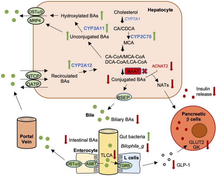
BA and glucose metabolism and gut dysbiosis in Baat −/− mice. Baat gene deletion reduces conjugated bile acids and increases unconjugated BAs dramatically in hepatocytes. Although BA excretion to the gallbladder is reduced due to higher affinity for conjugated BAs of bile salt export pump (BSEP), genes encoding basolateral BA export pumps (multidrug resistance‐associated protein 4 [MRP4] and OSTα/β) and hydroxylating enzymes such as CYP3A11, CYP2C70, and Cyp2A12 are increased in hepatocytes, which helps reduce accumulated unconjugated bile acid toxicity. Due to lower intestinal BA concentration, gut microbiota population is higher, but genus Bilophila, a sulfidogenic bacteria, is significantly less populated. Almost complete absence of TLCA fails to activate the Takeda G protein‐coupled receptor 5 (TGR5) signaling pathway and results in lower glucagon‐like peptide 1 (GLP‐1) level in serum. In addition, lower hepatic N‐acyl taurine levels due to absence of BAAT and lower synthesis of acyl‐CoA: amino acid N‐acyltransferases (ACNATs) lead to impaired glucose‐stimulated insulin secretion along with lower blood GLP‐1. ASBT, apical sodium–bile acid transporter; CDCA, chenodeoxycholic acid; GK, glucokinase; GLUT2, glucose transporter 2; NATs, N‐acyl taurines; NTCP, Na + −taurocholate co‐transporter peptide; OATP, organic anion transporting polypeptide.