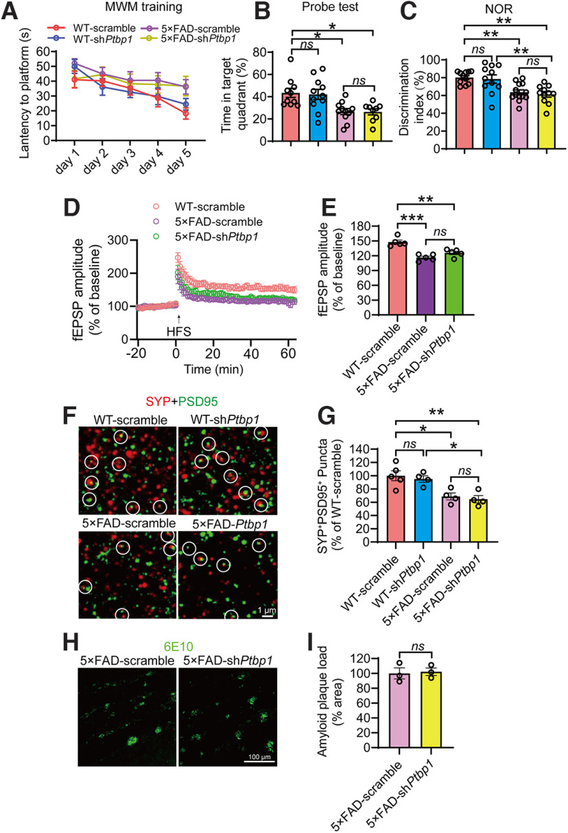
Figure 3.
Downregulation of PTBP1 in the hippocampus fails to attenuate cognitive and synaptic deficits as well as Aβ deposition in 5×FAD mice. A, B, MWM analysis of 5×FAD and WT control mice 2.5 months after shPtbp1 or the scramble control AAV transduction. The latency to find the hidden platform during the training phase (A), time spent in the target quadrant in the probe test (B); n = 9–11 animals, one-way ANOVA with Tukey's multiple comparisons. C, Quantification of discrimination indexes in the NOR test of the experimental mouse; n = 11–13 animals, one-way ANOVA with Tukey's multiple comparisons. D, E, Electrophysiological analysis of LTP. Recordings of hippocampal LTP induced by high-frequency stimulation (HFS, D), quantification of the field excitatory postsynaptic potentials (fEPSPs) during the last 10 min of LTP recording (E); n = 5 brain slices from 4 animals per group, one-way ANOVA with Tukey's multiple comparisons. F, G, Confocal analysis of SYP+PSD95+ synaptic puncta. Representative images (F), quantification (G); n = 4–5 animals, one-way ANOVA with Tukey's multiple comparisons. H, I, Confocal analysis of 6E10 (Aβ antibody) stained amyloid deposits. Representative images (H), quantification (I); n = 3 animals per group, unpaired t test. All quantified data are represented as mean ± SEM; *p < 0.05, **p < 0.01, ***p < 0.001. ns, Not significant.

