Figure 1.

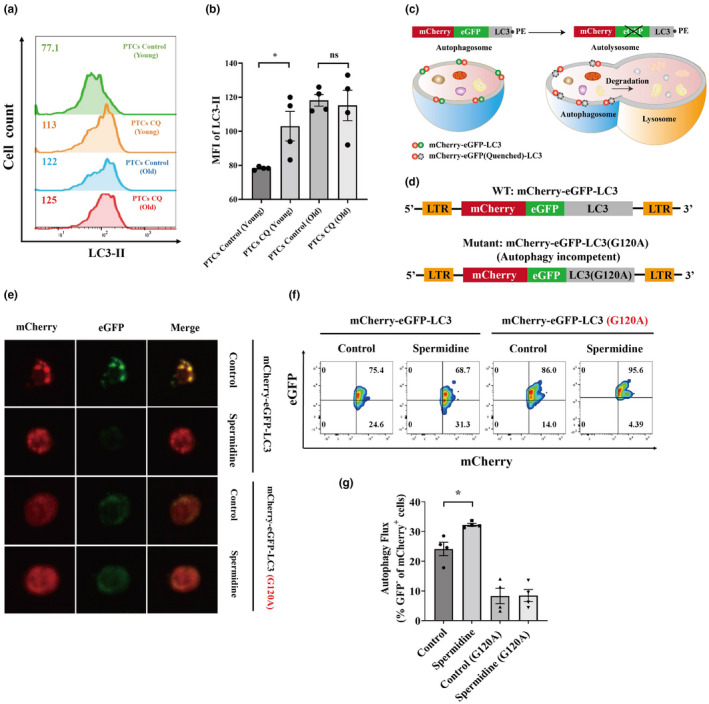
Autophagic flux impairment and restoration of senescent TCR‐Ts. (a, b) Peripheral T cells (PTCs) from young (25–30 years old) and old (more than 60 years old) donors were treated with or without chloroquine (CQ) (50 μm; 12 h), and LC3‐II was detected by flow cytometry. Representative flow cytometric results of LC3‐II in PTCs from young and old donors treated with or without CQ are shown (a). The LC3‐II expression levels in PTCs from young and old donors after CQ treatment are summarised and shown. Data represent mean ± SEM of n = 4 biological replicates (b). (c) Diagram showing the reporting mechanism of the mCherry‐GFP‐LC3 fusion protein. (d) Illustration of the lentiviral constructs encoding the autophagy gene LC3 in conjunction with mCherry and eGFP. Replacement of the LC3b glycine at the amino acid 120 position with alanine was used as an autophagy‐incompetent construct. (e) Representative confocal images defining the GFP and mCherry puncta in PTCs from old donors treated under the indicated conditions. (f, g) Representative flow cytometry plot (f) and quantification (g) of autophagic flux in the indicated conditions by measuring the loss of GFP in mCherry populations. Data represent mean ± SEM of n = 4 biological replicates. *P < 0.05; ns, not significant, the paired t‐test.
