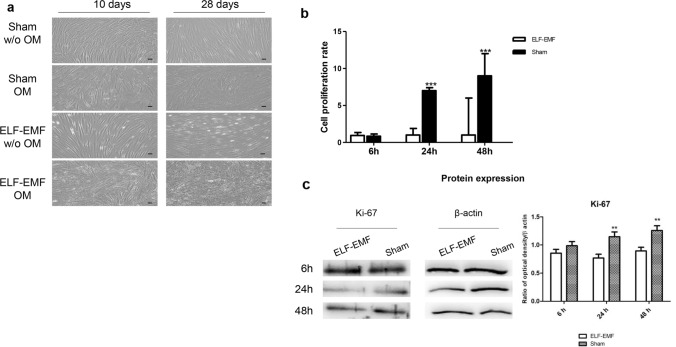
Fig. 4.
a Microphotographs of hPDLSCs cultured in the presence of experimental conditions for 10 and 28 days. Images were obtained using an inverted microscope (Leica DMi1, Wetzlar, Germany). Scale bar: 20 µm. b MTT assay was performed on hPDLSCs in presence of no-OM, and exposed to ELF-EMF or sham system, for 6, 24 and 48 h. c Western blot analysis of Ki-67 expression. Data are expressed as mean ± SD of two experiments performed in triplicate. ***p < 0.001

