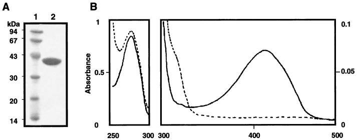
FIG. 2.
SDS-PAGE gel (A) and absorption spectra (B) of purified S. pombe Alr1p. (A) Lane 1, marker proteins (the molecular masses are shown in kilodaltons); lane 2, purified S. pombe Alr1p (10 μg). Protein was stained with Coomassie brilliant blue R-250. (B) The spectrum of the enzyme solution (1.0 mg/ml) in a 20 mM potassium phosphate buffer (pH 7.8) containing 0.1% 2-mercaptoethanol is shown as a solid line, while that of the enzyme dialyzed against 500 volumes of a 20 mM potassium phosphate buffer (pH 7.8) containing 0.1% 2-mercaptoethanol and 10 mM sodium borohydride is shown as a dashed line.

