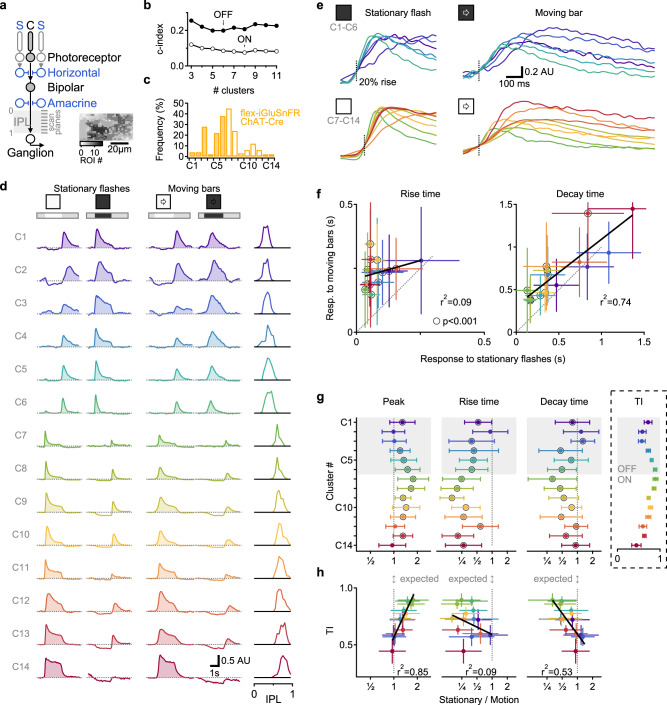
Fig. 1. Multiplexed representation of static and moving objects in BCs.
a Schematic of the investigated retinal circuits. Glutamate release in the IPL is predominantly mediated by bipolar cells (BCs). BCs integrate photoreceptor drive in their RF center (C) with an antagonistic surround (S) formed by horizontal and amacrine cells. Inset, exemplar ROIs identified from iGluSnFR fluorescence in a single scan plane. b Diversity of responses to stationary flashes from 1828 ROIs (278 scan fields in 67 animals, 34 females; ages p43-359), arrows indicate the optimal number of functional clusters of glutamate release within the shown range. c Distribution of the clusters imaged from floxed-iGluSnFR expressed under the ChAT-Cre promotor. d Mean clusters’ responses, sorted by pixel depth distribution in the IPL (right). e Focus on the rising phase of the signals. f Mean (±SD) clusters’ kinetics, linear fits in black. Clusters with a significant (p < 0.001; paired t test) difference between motion vs. stationary response are indicated by black circles. g, h IPL depth (g) or transiency index (TI; h, inset in g) vs. the mean (±SD) ratio between the stationary and motion responses for each of the glutamate clusters. Color coding in e–h by cluster identity. Black circles, clusters with stationary/motion response ratio with significant (p < 0.001, t test) difference from unity.

