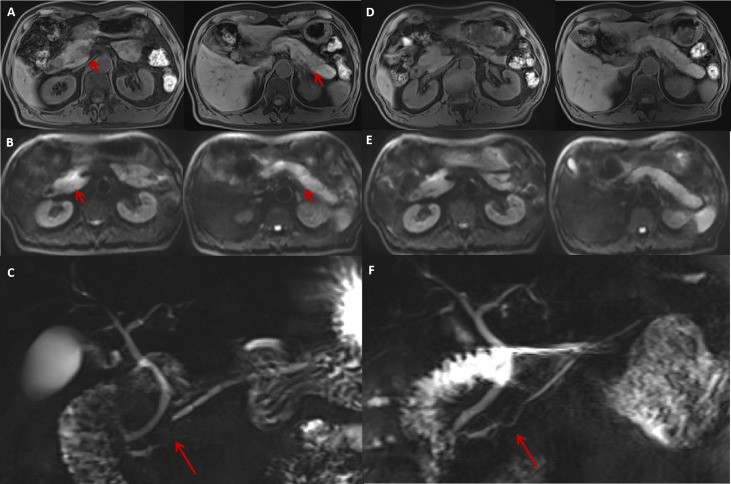
Figure 1.
The MRI features of irAP and its improvement after treatment. The MRI at diagnosis revealed a slightly swollen pancreas with multiple nodular (A, arrow) and patchy abnormal signals with decreased intensity on T1WI sequences (A) and increased intensity on DWI (B, arrow) located in the pancreatic head and tail. MRCP showed segmental stenosis of the MPD in the pancreatic head (C, arrow). After 1 month of prednisolone treatment, the swelling reduced and the abnormal signals decreased on T1W1 (D) and DWI (E) sequences. The MPD stenosis also resolved (F, arrow). irAP, immune-related acute pancreatitis; MRI, magnetic resonance imaging; T1W1, T1-weighted image; DWI, diffusion-weighted imaging; MPD, main pancreatic duct; MRCP, magnetic resonance cholangiopancreatography.

