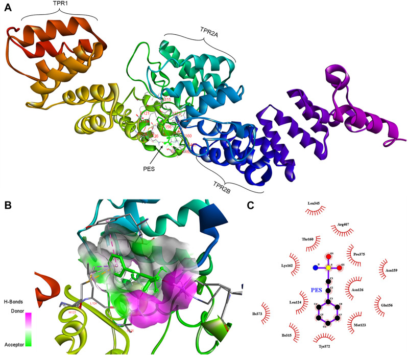
FIGURE 3.
Schematic representing the interaction of PES with PfHop. (A) A docked three-dimensional model with a ribbon structure of full-length PfHop bound to PES. The domains of the protein are also shown. PES binds to the TPR1, DP1, and TPR2B regions of the protein. (B) Surface and ribbon views of the docked structures, showing the residues involved in the interaction. (C) A 2D illustration of the receptor-ligand interface. PES appears to hydrogen bond with residues Arg407 on PfHop. Thus, PES forms pi-pi T-shaped interactions with Tyr372, pi-alkyl with Leu124 and Ile373, pi sigma, and pi-sulfur with Met123 of PfHop.

