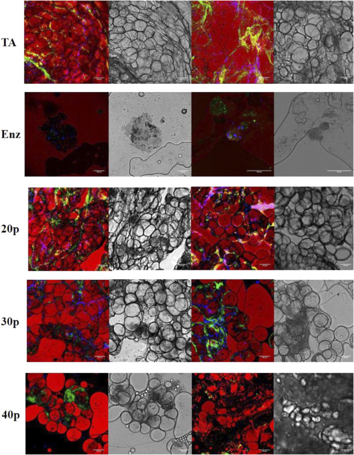
FIGURE 6.
Immunofluorescence by confocal microscopy of the different SVFs. In red, adipocytes are stained by BODIPY; in green, endothelial cells are stained by isolectin coupled with Alexa Fluor 488, and in blue, nucleated cells are stained by DAPI. Shades of gray: transmission electron microscopy imaging of the same section. * False positive, lipid vacuole. SVF: stromal vascular fraction.

