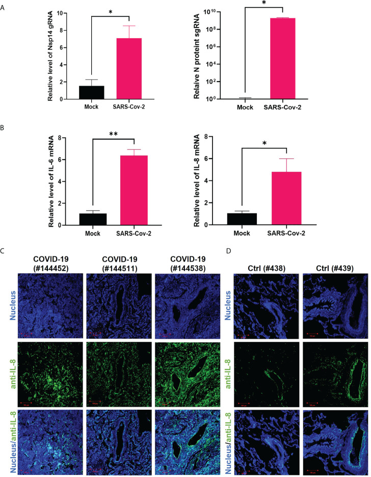
Figure 3.
IL-8 is induced by Nsp14 and in SARS-CoV-2 infected lung tissues. (A, B) HEK293T-ACE2 cells were infected with wild-type SARS-Cov-2 viruses. Cells were harvested at 24 h. Total RNAs were extracted, and the expression of viral genes (Nsp14, N-protein, A) or cytokines (IL-6, IL-8, B) was analyzed by RT-qPCR and normalized to mock infection. Results were calculated from 3 technical repeats and presented as mean +/- standard error of the mean (SEM). (* p<0.05; ** p<0.01; by unpaired Student’s t-test). (C, D) Dissected lung tissues from COVID19 patients (C, donors #144452, #144511, #144538) or non-infected donors (D, donors #438, #439) were analyzed for IL-8 expression by immunofluorescence (green). Nuclei were stained with Hoechst (blue). Scale bar: 100 µm.

