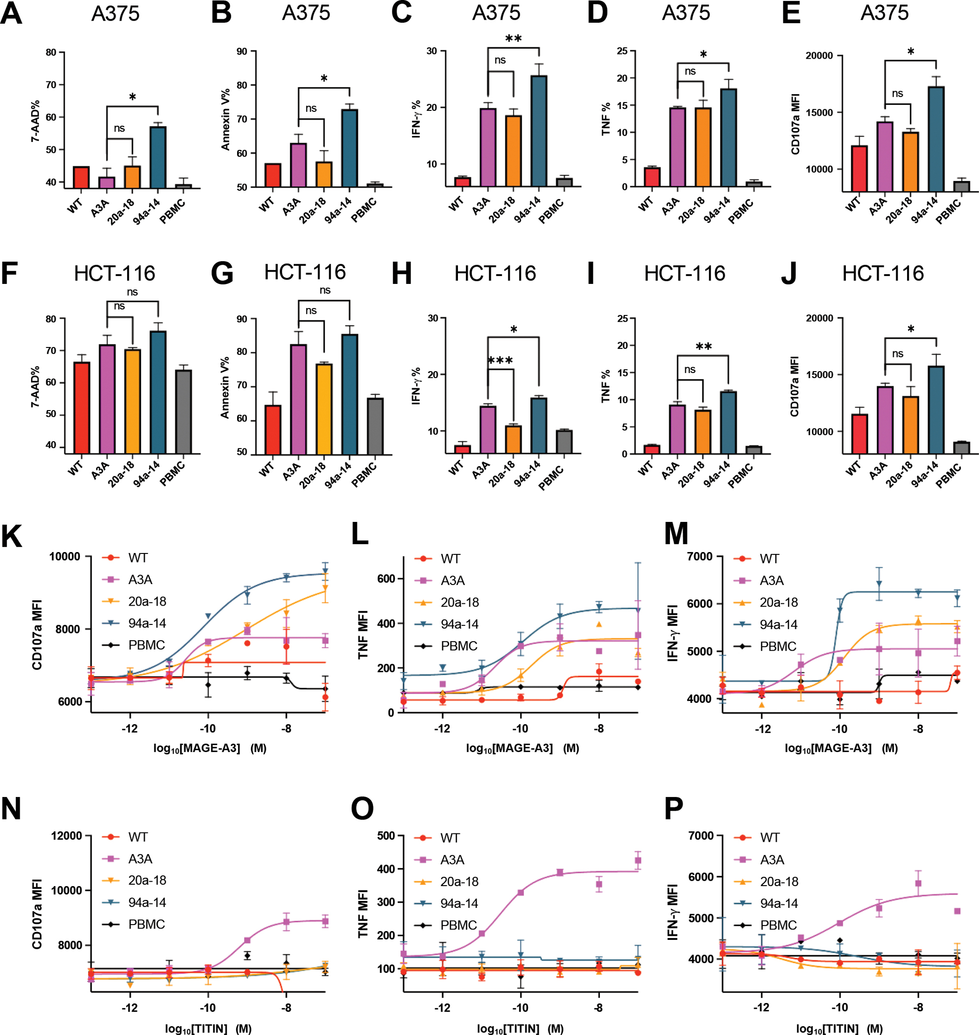
Figure 5. Cytotoxicity and specificity of engineered MAGE-A3-specific TCR.

(A-B) Killing of A375 melanoma cell line by different MAGE-A3-specific TCR transduced human primary T cells.
(C-E) IFN-γ, TNF, and cytotoxic granule release (CD107a staining) by different MAGE-A3-specific TCR transduced human primary T cells, induced by the A375 melanoma cell line.
(F-G) Killing of HCT-116 colon cancer cell line by different MAGE-A3-specific TCR transduced human primary T cells.
(H-J) IFN-γ, TNF, and cytotoxic granule release (CD107a staining) by different MAGE-A3-specific TCR transduced human primary T cells, induced by the HCT-116 colon cancer cell line.
(K-M) Cytotoxic granule release (CD107a staining), TNF, and IFN-γ by different MAGE-A3-specific TCR transduced human primary T cells, induced by HLA-A1+ 293T cells pulsed with a titration of MAGE-3 peptide.
(N-P) Cytotoxic granule release (CD107a staining), TNF, and IFN-γ by different MAGE-A3-specific TCR transduced human primary T cells, induced by HLA-A1+ 293T cells pulsed with a titration of TITIN peptide.
(A-P) Data are representative of 3 independent experiments. Data are shown as mean ± SD of technical duplicates. ns: not significant; ✱: P<0.05; ✱✱: P<0.01; ✱✱✱: P<0.001; ✱✱✱✱: P<0.0001
