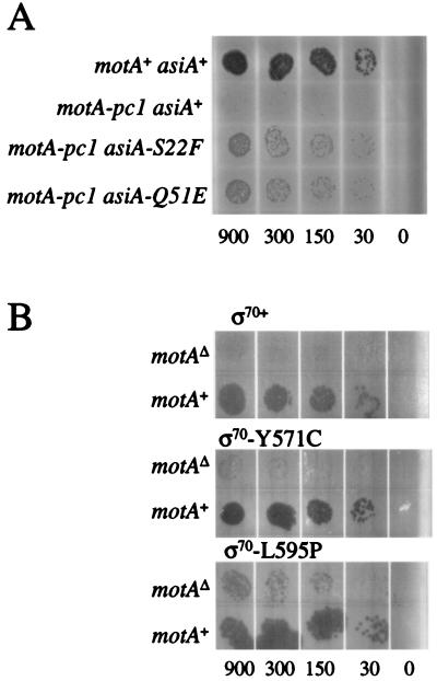
FIG. 2.
Plaque spot tests to visualize suppression. (A) The plate contained a lawn of TabG pBSPLO+ cells, and the indicated T4 K10 strain was spotted on the lawn in 3-μl aliquots. The aliquots contained approximately the number of phage indicated below the plate. The positive control was T4 strain K10 (motA+ asiA+), which resulted in full growth (top row), while the negative control K10 motA-pc1 (asiA+) showed very little growth (second row). (B) The plates contained lawns of TabG cells with the indicated ς70 expression plasmid and a MotA-pc1 expression plasmid. Phage T4 denAB motAΔ (top row) or T4 denAB (motA+; bottom row) were spotted on the lawn in 3-μl aliquots that contained approximately the number of phage indicated below the plate. All plates were incubated at 37°C overnight.

