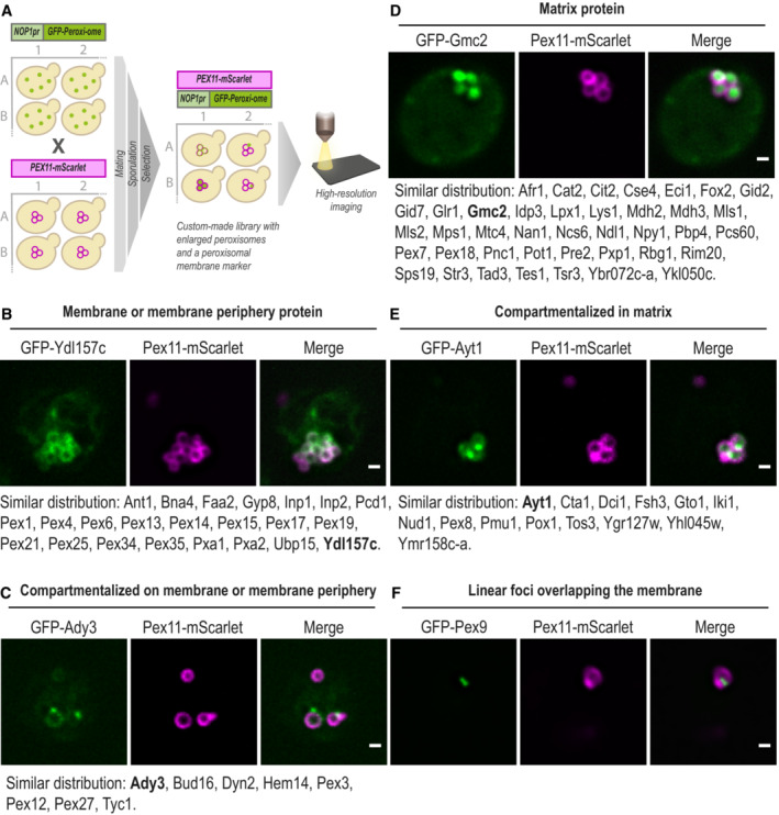
Figure 2. High‐resolution imaging reveals the sub‐organellar distribution of the peroxi‐ome.

- The sub‐peroxisomal localization of each peroxi‐ome protein was captured by high‐resolution imaging and was enabled by genomic and metabolic enlargement of peroxisomes. Enlargement was induced by the integration of the C′ tagged form of the peroxisomal‐membrane protein Pex11 into a yeast N′ GFP collection using an automated mating procedure and upon 20 h of incubation in oleate‐containing media.
- GFP‐Ydl157c represents a protein that distributes homogeneously on the peroxisomal membrane or membrane periphery.
- GFP‐Ady3 represents a protein that compartmentalizes on the peroxisomal membrane or membrane periphery.
- GFP‐Gmc2 represents a protein that distributes homogeneously in the peroxisomal matrix.
- GFP‐Ayt1 represents a protein that compartmentalizes in the matrix.
- GFP‐Pex9 was the only protein that showed linear foci overlapping the peroxisomal membrane.
Data information: For each category, proteins with similar distributions are listed below the micrographs, when the protein in bold is the one presented. For all micrographs, a single focal plane is shown. The scale bar is 500 nm.
