
Figure 2.
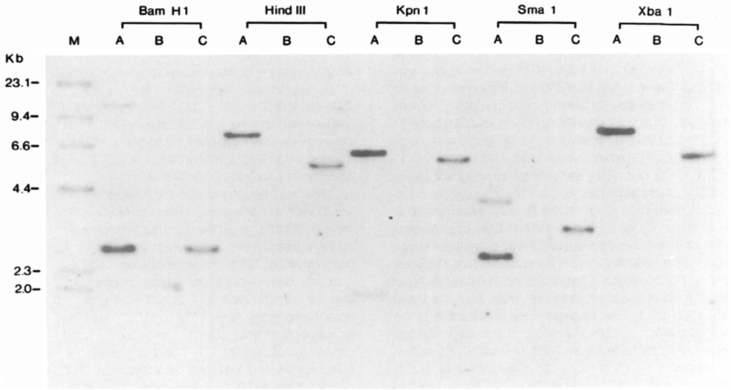
Southern Blot Analysis of Integrated HIV Envelope Sequences in CEM Cells
CEM cells were infected with retroviral env and tat expression vectors by cocultivation with DNA transfected amphotrophic packaging cell line PA317 in the presence of polybrene. Infected cells were selected in G418 and analyzed for envelope sequences. a, CEM cells infected with pDOL CMV·env; b, uninfected CEM cells; c, CEM cells infected with pDOL·tat. Genomic DNA was prepared by guanidine thiocyanate lysis, restriction endonuclease digested, electrophoresed, and transferred to nylon membranes. Blots were hybridized under high stringency conditions to a 32P-labeled probe prepared by nick translation of a Sall–Xhol fragment of λ-N1G-F. Molecular weight markers are provided by 32P end-labeled HindIII digests of λDNA.