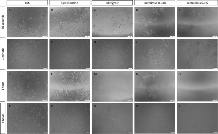
Figure 5.
Immortalized human corneal epithelial cells morphology following anti-inflammatory treatments. Brightfield images of iHCE cells 72 hours following treatments with saline (A, F, K, P), cyclosporine (B, G, L, Q), lifitegrast (C, H, M, R), tacrolimus 0.03% (D, I, N, S), and tacrolimus 0.1% (E, J, O, T). Treatment times: 30 seconds (A–E), 1 minute (F–J), 1 hour (K–O), and 4 hours (P–T). Cells treated with 20% tested drug, 80% normal growth medium. Scale bar: 200 µm.

