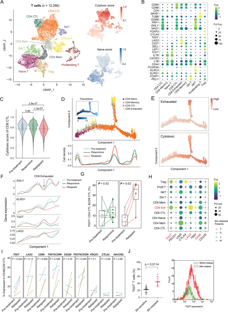
Fig. 2.
Elevated levels of cytotoxic T cells overexpressing TIGIT post relapse. (A) Combined UMAP plots of all T-cell subsets. Each dot indicates an individual cell; color denotes T-cell subsets (left), cytotoxic score, and naïve score (right). (B) Bubble heatmap showing marker genes across T cell clusters from A. Dot size indicates fraction of expressing cells, colored according to normalized expression levels. (C) Boxplots showing the distribution of cytotoxic score of CD8+ CTL cells. Mann-Whitney test used to calculate the significances. (D) Top, Monocle2 trajectory plot of CD8+ T cells. Cell orders are inferred from expression of most differential genes across CD8+ T-cell subpopulations. Color is coded by CD8+ T-cell subpopulations. Insert visualizes the pseudotime defined by Monocle2. Bottom, cell density relevant to BA response along with component 1 of Monocle2 trajectory. (F) Average gene expression of cytotoxic markers and exhaustion markers along with component 1 of Monocle2 trajectory. Loess regression lines of each gene’s expression are shown. (G) Pairwise comparison of the fraction of combined CTLs (CD4+ and CD8+) expressing TIGIT among T cells for pre- vs post-treatment samples at the responsive stage (left) or post relapse (right). (H) Bubble heatmap showing immune checkpoint molecules across T-cell clusters from (A). Dot size indicates fraction of expressing cells, colored according to normalized expression levels. (I) Pairwise comparison of the fraction of combined CTLs (CD4+ & CD8+) expressing immune checkpoint molecules for pre- vs post-treatment samples post relapse. (J) TIGIT expression is upregulated on the cell surface of T cells in the tumor microenvironment of BA-relapsed patients (n = 4) compared to BA-sensitive patients (n = 7) (left panel). TIGIT expression on T cells was assessed after relapse compared to before relapse in a representative patient (right panel)

