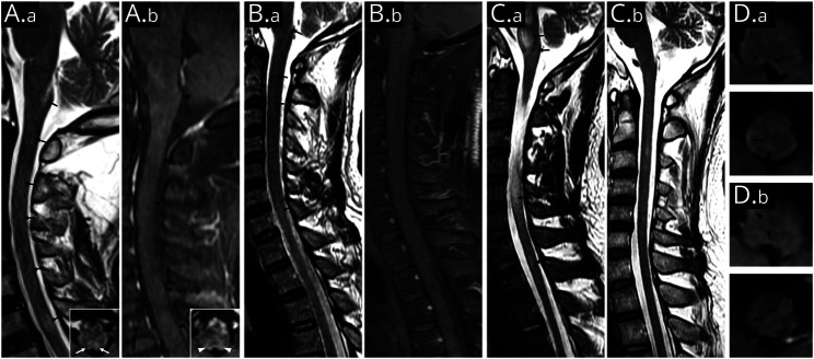
Figure 3. Area Postrema and Dorsal Medulla Lesions in Representative Glial Fibrillary Acidic Protein-IgG–Positive Patients.
Sagittal T2-weighted fluid-attenuated inversion recovery scan shows lesions in the area postrema and multifocal punctate lesions in medulla and cervical spinal cord (A.a [patient 5] and B.a [patient 1]; black arrows). Inset in (A.a) is the axial image (area postrema level), which shows bilateral lesions involving the dorsal medulla (white arrows). Sagittal T1-weighted MRI with gadolinium shows irregular patchy or linear enhancement in the medulla and cervical spinal cord (A.b and B.b; black arrowheads). Inset in (A.b) is the axial image (area postrema level), which shows hazy gadolinium enhancement of bilateral dorsal medulla (white arrowheads). T2-hyperintensity lesions in pons, medulla oblongata, and cervical longitudinally extensive lesions were noted at the initial presentation of patient 4 (C.a). Follow-up MRI at 6 months after immunotherapy reveals remission of most lesions (C.b). Axial images of patient 2 show scattered faint T2-hyperintensity lesions in dorsal medulla oblongata (D.a; black arrows) accompanied by patchy gadolinium enhancement (D.b; black arrowheads).

