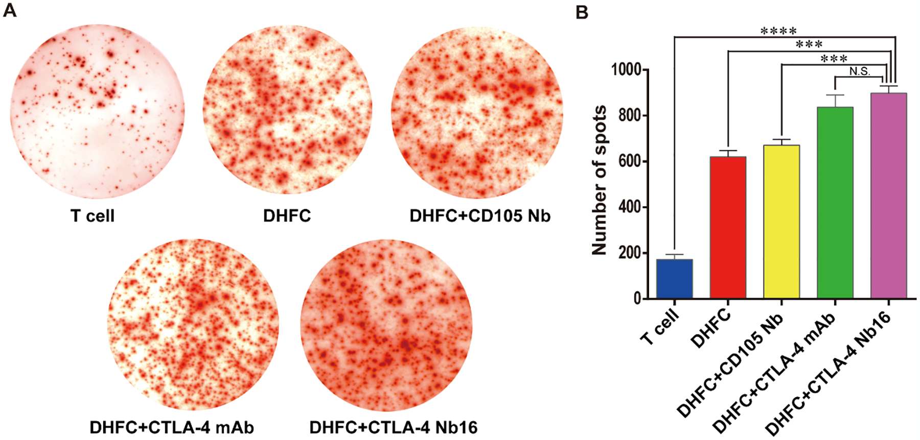
Figure 3. CTLA-4 Nb16 increased the abundance of IFN-γ secreting CD8+ T lymphocytes.

(A) CD8+ cells were mixed with different antibodies following with DHFC, and then co-cultured for 7 days. ELISPOT assay was applied to examine the number of CD8+ T lymphocytes secreting IFN-γ. (B) The number of spots in DHFC+CTLA-4 Nb16 group increased significantly than other’s groups, except DHFC+CTLA-4 mAb groups. Data are means ± SD, n = 3. *** P < 0.001, **** P < 0.0001.
