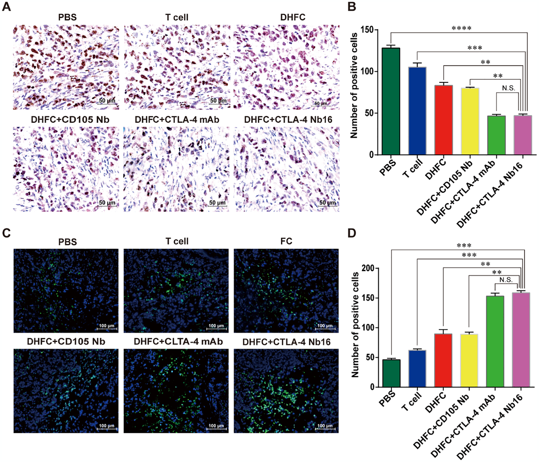
Figure 6. CTLA-4 Nb16 stimulation suppressed proliferation of tumor cells and promoted tumor cell apoptosis in mice.

(A) Immunohistochemical assay of Ki67 staining in different groups (x 40). Brown in nuclear indicates the positive staining. (B) The Ki67 expression of tumor cells in DHFC+CTLA4-Nb16 group was significant lower than in other groups, except in DHFC+CTLA4-mAb group. (C) TUNEL assay was used to monitor cell apoptosis in different groups. Blue indicates nuclear and green indicates the positive signal (×20). (D) The apoptosis of tumor cells in DHFC+CTLA4-Nb16 group was significant higher than that in other’s groups, except in DHFC+CTLA4-mAb group. Data are measn ± SD. **P < 0.01, *** P < 0.001, **** P < 0.0001.
