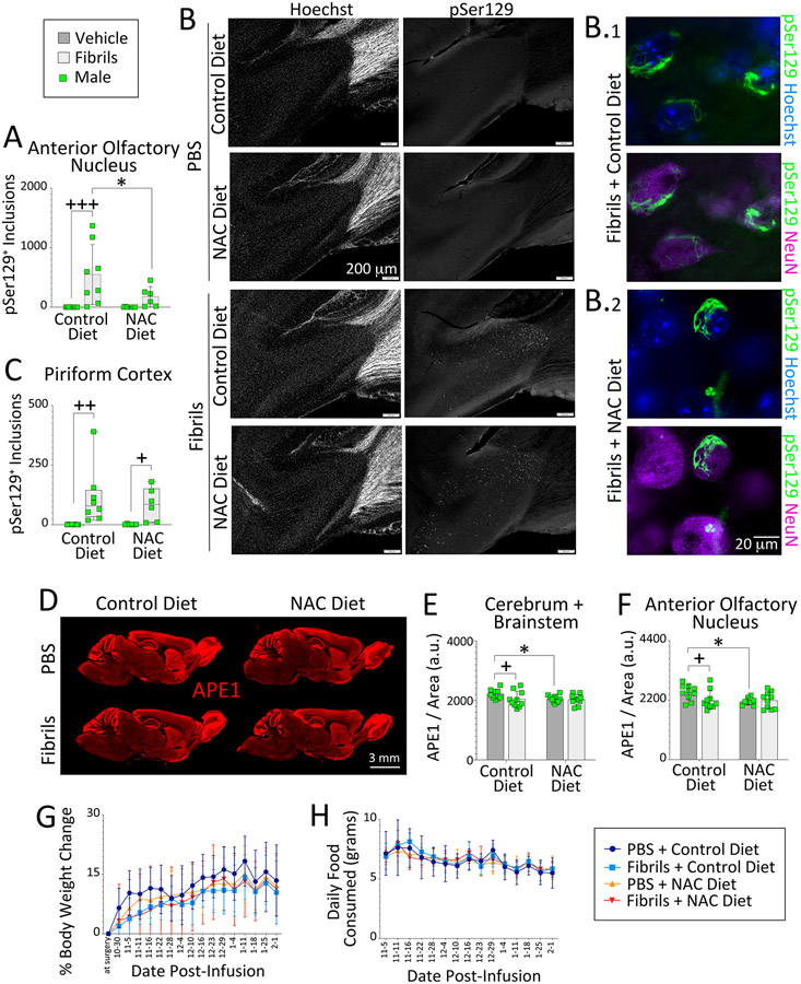
Figure 6. Oxidative stress influences APE1 expression in male mice.
Four-month-old male mice were infused in the right OB/AON with sonicated α-synuclein fibrils (5 μg) or PBS (1 μL). Mouse chow with and without N-acetylcysteine (~200 mg NAC/kg bodyweight) was administered for three months following fibril or PBS infusions. Sagittal brain sections were collected after a three-month survival period at 7 months of age and immunostained for pSer129 and APE1. (A) A blinded observer counted pSer129+ structures per field of view in the (A-B) AON and (C) piriform cortex of the right hemisphere. Higher magnification images of inclusions in the AON of fibril-infused animals are shown in B.1-2, vis-à-vis nuclear staining with NeuN antibodies or the Hoechst reagent (also see Fig. S14-S15). (D-F) A blinded observer analyzed APE1 expression levels in the (E) cerebrum and brainstem, as well as in the (F) AON of the right hemisphere by tracing the regions of interest on (D) low-resolution, high-sensitivity scans captured with a 16 bit-depth imager. Line graphs showing (G) % bodyweight change and (H) daily food consumption as a function of days post-infusion. Data in panels A and E-F are shown as the mean ± S.D. Two-way ANOVAs were followed by the Bonferroni post hoc. Data in C are shown are box plots with interquartile ranges, analyzed by the Kruskal-Wallis test and Dunn post hoc. *p≤0.05 Control vs. NAC diet; + p≤0.05, ++ p≤0.01, +++p≤0.001 PBS vs. fibrils.

