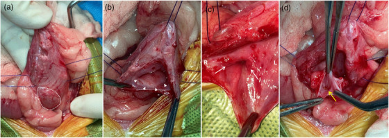
FIGURE 2.

Intraoperative photographs of the urinary bladder with a ureterocele (a–d). (a) The bladder wall is swollen outward (white dotted line) at the ureterovesical junction. (b, c) The cystic structure (white arrowheads) within the bladder wall is seen, and both sides of the ureteral orifice (white arrows) are at the normal position within the trigone. (d) Several small calculi (yellow arrow) can be seen through the incision made on the mucosal surface of the ureterocele. R, right; L, left
