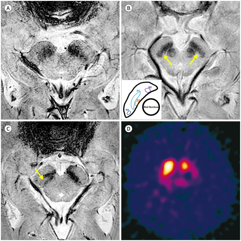
Fig. 2. Nigrosome imaging in Parkinson’s disease.
A. 3T SWI from a 65-year-old male patient diagnosed with Parkinson’s disease shows loss of nigral hyperintensity created by the nigrosome 1.
B. An 85-year-old female patient shows bilateral loss of nigrosome 1 hyperintensity upon susceptibility-map weighted imaging, but the hyperintense presumed nigrosome 4 (arrows) at the level of the lower red nucleus that is medial to the nigrosome 1 can be identified.
C, D. 3T SWI from a 75-year-old female patient shows intact nigral hyperintensity in the right side (arrow) and loss of nigral hyperintensity in the left side, which is correlated with her unilateral right-side motor symptom. Her 123I-2β-carbomethoxy-3β-(4-iodophenyl)-N-(3-fluoropropyl)-nortropane single photon emission computed tomography (123I-FP-CIT SPECT) shows the corresponding asymmetric dopamine transporter uptake in the left striatum.
SWI = susceptibility-weighted imaging

