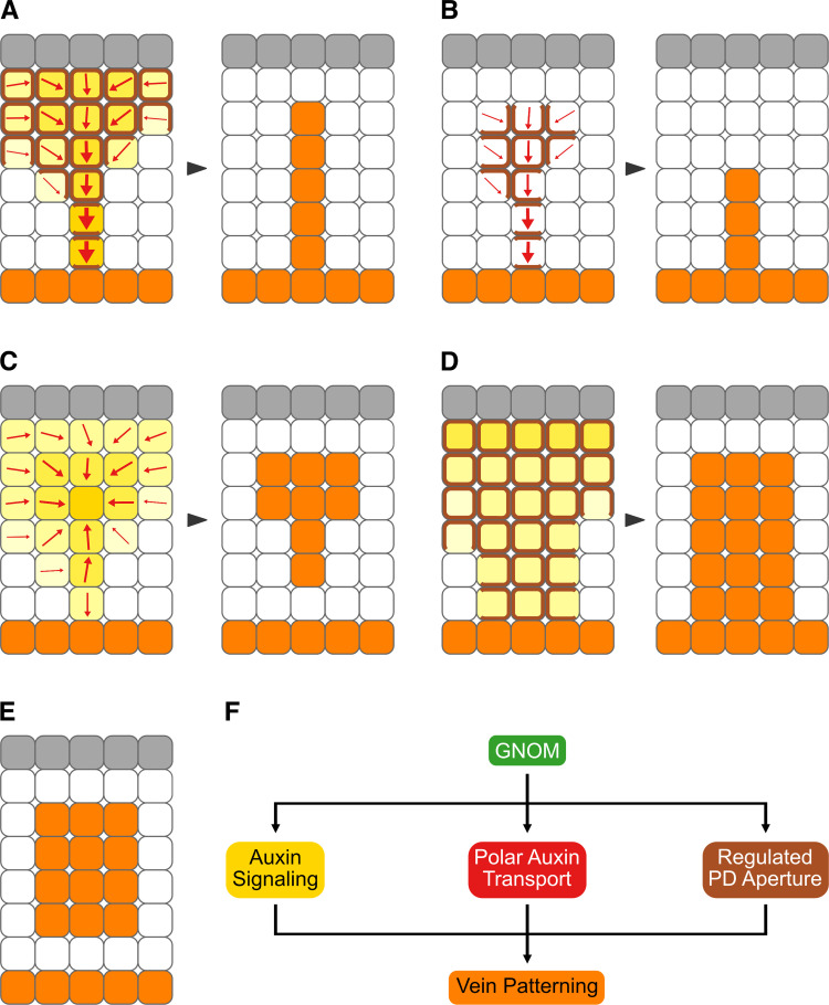
Fig 7. Summary and interpretation.
(A–F) Gray: epidermis, whose role in vein patterning—if any—remains unclear [147]. Increasingly darker yellow: progressively stronger auxin signaling. Increasingly thicker arrows: progressively more polarized auxin transport. Brown: PD-mediated cell–cell connection. Orange: veins. Arrowheads temporally connect vein patterning stages with mature vein patterns. (A) In WT, veins are patterned by gradual restriction of auxin signaling domains [65,67,88,125,126,148], gradual restriction of auxin transport domains and polarization of auxin transport paths [5,37,38,56,65–67,126,148], and gradual reduction of PD permeability between incipient veins and surrounding nonvascular tissues. (B) Inhibition of auxin signaling leads to narrower domains of auxin transport [5,65,148] and promotes reduction of PD permeability between incipient veins and surrounding nonvascular tissues. (C) Defects in the ability to regulate PD aperture lead to weaker and broader domains of auxin signaling, fragmentation of auxin transport domains, and abnormal polarization of auxin transport paths. (D) Inhibition of auxin transport leads to weaker and broader domains of auxin signaling [5,38,48,148] and delays reduction of PD permeability between incipient veins and surrounding nonvascular tissues. (E) Loss of GN function or simultaneous inhibition of auxin signaling, polar auxin transport, and ability to regulate PD aperture leads to clusters of vascular cells. (F) Veins are patterned by the coordinated activities of three GN-dependent pathways: auxin signaling, polar auxin transport, and regulated PD aperture. GN, GNOM; PD, plasmodesma; WT, wild-type.

